Astaraki Mehdi, Severgnini Mara, Milan Vittorino, Schiattarella Anna, Ciriello Francesca, de Denaro Mario, Beorchia Aulo, Aslian Hossein
Department of Biomedical Engineering and Health Systems, KTH Royal Institute of Technology, Sweden.
Department of Medical Physics, Azienda Sanitaria Universitaria Integrata di Trieste, Trieste, Italy.
Phys Imaging Radiat Oncol. 2018 Mar 5;5:52-57. doi: 10.1016/j.phro.2018.02.003. eCollection 2018 Jan.
In radiation therapy, defining the precise borders of cancerous tissues and adjacent normal organs has a significant effect on the therapy outcome. Deformable models offer a unique and robust approach to medical image segmentation. The objective of this study was to investigate the reliability of segmenting organs-at-risk (OARs) using three well-known local region-based level-set techniques.
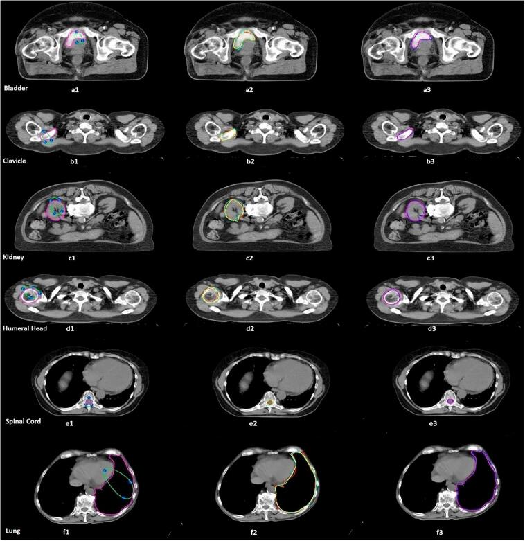
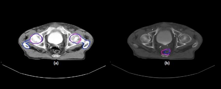
A total of 1340 non-enhanced and enhanced planning computed tomography (CT) slices of eight OARs (the bladder, rectum, kidney, clavicle, humeral head, femoral head, spinal cord, and lung) were segmented by using local region-based active contour, local Chan-Vese, and local Gaussian distribution models. Quantitative metrics, namely Hausdorff Distance (HD), Mean Absolute Distance (MAD), Dice coefficient (DC), Percentage Volume Difference (PVD) and Absolute Volumetric Difference (AVD), were adopted to measure the correspondence between detected contours and the manual references drawn by experts.
The results showed the feasibility of using local region-based active contour methods for defining six of the OARs (the bladder, kidney, clavicle, humeral head, spinal cord, and lung) when adequate intensity information is available. While the most accurate results were achieved for lung (DC = 0.94) and humeral head (DC = 0.92), a poor level of agreement (DC < 0.7) was obtained for both rectum and femur.
Incorporating local statistical information in level set methods yields to satisfactory results of OARs delineation when adequate intensity information exists between the organs. However, the complexity of adjacent organs and the lack of distinct boundaries would result in a considerable segmentation error.
在放射治疗中,确定癌组织和相邻正常器官的精确边界对治疗结果有重大影响。可变形模型为医学图像分割提供了一种独特且强大的方法。本研究的目的是使用三种著名的基于局部区域的水平集技术来研究分割危及器官(OARs)的可靠性。
使用基于局部区域的活动轮廓、局部Chan-Vese和局部高斯分布模型,对八个OARs(膀胱、直肠、肾脏、锁骨、肱骨头、股骨头、脊髓和肺)的总共1340张非增强和增强的计划计算机断层扫描(CT)切片进行分割。采用定量指标,即豪斯多夫距离(HD)、平均绝对距离(MAD)、骰子系数(DC)、体积百分比差异(PVD)和绝对体积差异(AVD),来测量检测到的轮廓与专家绘制的手动参考之间的对应关系。
结果表明,当有足够的强度信息时,使用基于局部区域的活动轮廓方法来定义六个OARs(膀胱、肾脏、锁骨、肱骨头、脊髓和肺)是可行的。虽然肺(DC = 0.94)和肱骨头(DC = 0.92)取得了最准确的结果,但直肠和股骨的一致性水平较差(DC < 0.7)。
当器官之间存在足够的强度信息时,在水平集方法中纳入局部统计信息会产生令人满意的OARs描绘结果。然而,相邻器官的复杂性和缺乏明显边界会导致相当大的分割误差。