Houri Jordan, Karunamuni Roshan, Connor Michael, Pettersson Niclas, McDonald Carrie, Farid Nikdokht, White Nathan, Dale Anders, Hattangadi-Gluth Jona A, Moiseenko Vitali
Department of Radiation Medicine and Applied Sciences, University of California San Diego, La Jolla, CA, USA.
Department of Physics, University of Oxford, Oxford, UK.
Phys Imaging Radiat Oncol. 2018 May 1;6:39-46. doi: 10.1016/j.phro.2018.04.003. eCollection 2018 Apr.
Brain radiotherapy (RT) can cause white matter damage and downstream neurocognitive decline. We developed a computational neuroimaging tool to regionally partition individual white matter tracts, then analyze regional changes in diffusion metrics of white matter damage following brain RT.
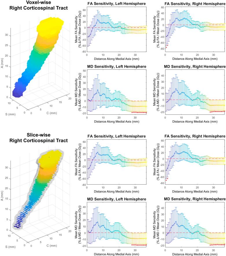
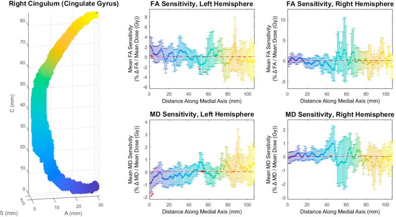
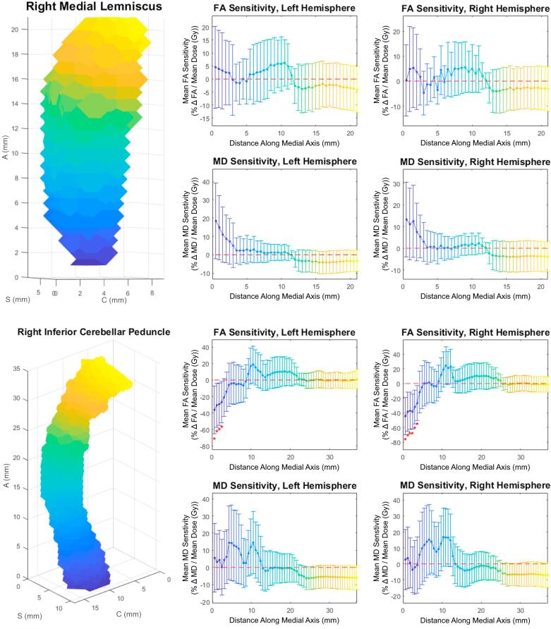
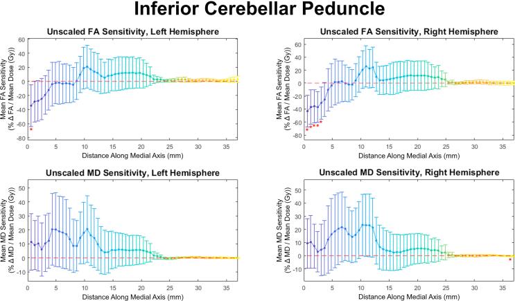
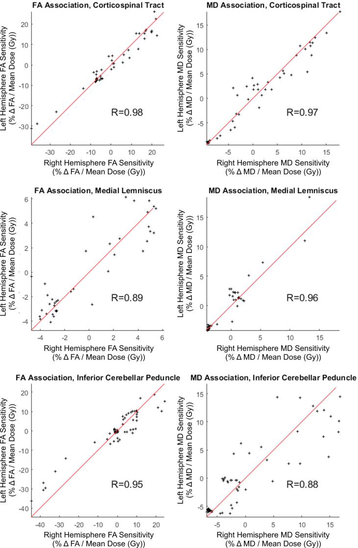
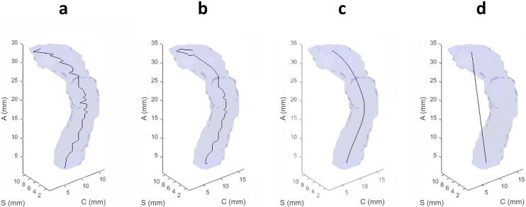
RT dose, diffusion metrics and white matter tract structures were extracted and mapped to a reference brain for 49 patients who received brain RT, and underwent diffusion tensor imaging pre- and 9-12 months post-RT. Based on their elongation, 23 of 48 white matter tracts were selected. The Tract-Crawler software was developed in MATLAB to create cross-sectional slice planes normal to a tract's computed medial axis. We then performed slice- and voxel-wise analysis of radiosensitivity, defined as percent change in mean diffusivity (MD) and fractional anisotropy (FA) as a function of dose relative to baseline.
Distinct patterns of FA/MD radiosensitivity were seen for specific tracts, including the corticospinal tract, medial lemniscus, and inferior cerebellar peduncle, in particular at terminal ends. These patterns persisted for corresponding tracts in left and right hemispheres. Local sensitivities were as high as 40%/Gy (e.g., voxel-wise: -39 ± 31%/Gy in right corticospinal tract FA, -45 ± 25%/Gy in right inferior cerebellar peduncle FA), p < 0.05.
Tract-Crawler, a novel tool to visualize and analyze cuts of white matter structures normal to medial axes, was used to demonstrate that particular white matter tracts exhibit significant regional variations in radiosensitivity based on diffusion biomarkers.
脑部放射治疗(RT)可导致白质损伤及下游神经认知功能衰退。我们开发了一种计算神经成像工具,用于对个体白质束进行区域划分,进而分析脑部放疗后白质损伤扩散指标的区域变化。
提取49例接受脑部放疗患者的放疗剂量、扩散指标和白质束结构,并将其映射到一个参考脑上,这些患者在放疗前及放疗后9 - 12个月接受了扩散张量成像。根据其伸长情况,从48条白质束中选取了23条。在MATLAB中开发了Tract - Crawler软件,以创建垂直于束计算中轴线的横截面切片平面。然后,我们对放射敏感性进行了切片和体素层面的分析,放射敏感性定义为平均扩散率(MD)和各向异性分数(FA)相对于基线随剂量变化的百分比。
在特定束中观察到FA/MD放射敏感性的不同模式,包括皮质脊髓束、内侧丘系和小脑下脚,特别是在末端。这些模式在左右半球的相应束中持续存在。局部敏感性高达40%/Gy(例如,体素层面:右侧皮质脊髓束FA为 - 39 ± 31%/Gy,右侧小脑下脚FA为 - 45 ± 25%/Gy),p < 0.05。
Tract - Crawler是一种用于可视化和分析垂直于中轴线的白质结构切片的新型工具,用于证明基于扩散生物标志物,特定白质束在放射敏感性上表现出显著的区域差异。