Smt Kanuri Santhamma Center for Vitreo-Retinal Diseases, L V Prasad Eye Institute, Hyderabad, Telangana, India.
Tej Kohli Cornea Institute, L V Prasad Eye Institute, Hyderabad, Telangana, India.
Indian J Ophthalmol. 2021 Feb;69(2):423-425. doi: 10.4103/ijo.IJO_1161_20.
Deeply embedded corneal foreign bodies and intrastromal foreign body removal can often be a challenge. The aim of this report was to describe the utility of endoscopy in visualization and removal of an embedded corneal bee stinger.
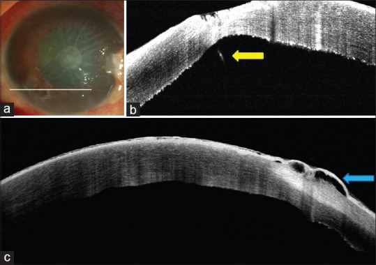
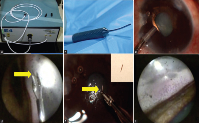
A 44-year-old male patient developed toxic keratopathy after injury from a bee stinger. On examination, the bee stinger was noted to be deeply embedded in the corneal stroma. A superficial keratectomy was initially attempted; however, the stinger was noted to be intrastromal and protruding into the anterior chamber and could not be removed. An Endoscopy-assisted visualization was used to remove the stinger.
The bee stinger was successfully removed and the patient's vision improved to 20/100 from an initial CFCF (counting fingers close to face) at time of presentation. At the end of 3 months follow-up, there was residual corneal edema along with cataractous changes in the lens as a sequelae of the initial bee sting injury. The patient subsequently underwent an endothelial keratoplasty along with phacoemulsification with intraocular lens implantation and the final BCVA improved to 20/40.
Endoscopyassisted visualisation of anterior chamber and angle structures can be valuable in removal of retained and deeply embedded corneal or intracameral foreign bodies.
深层角膜异物和基质内异物的取出常常具有挑战性。本报告的目的是描述内镜在可视化和取出嵌顿角膜蜜蜂蜇刺中的应用。
一名 44 岁男性患者因蜜蜂蜇伤后发生中毒性角膜炎。检查时发现蜜蜂蜇刺深深地嵌入角膜基质中。最初尝试进行浅层角膜切除术;然而,蜇刺位于基质内并突出到前房,无法取出。采用内镜辅助可视化技术取出蜇刺。
成功取出蜜蜂蜇刺,患者的视力从就诊时的初始近距手动(CF)改善至 20/100。在 3 个月的随访结束时,由于初始蜜蜂蜇伤的后遗症,仍存在角膜水肿和晶状体白内障改变。患者随后接受内皮角膜移植术联合超声乳化白内障吸除术和人工晶状体植入术,最终最佳矫正视力(BCVA)提高至 20/40。
内镜辅助可视化前房和房角结构可有助于取出残留和深层嵌入的角膜或眼内异物。