• 文献检索
  • 文档翻译
  • 深度研究
  • 学术资讯
  • Suppr Zotero 插件Zotero 插件
  • 邀请有礼
  • 套餐&价格
  • 历史记录
应用&插件
Suppr Zotero 插件Zotero 插件浏览器插件Mac 客户端Windows 客户端微信小程序
定价
高级版会员购买积分包购买API积分包
服务
文献检索文档翻译深度研究API 文档MCP 服务
关于我们
关于 Suppr公司介绍联系我们用户协议隐私条款
关注我们

Suppr 超能文献

核心技术专利:CN118964589B侵权必究
粤ICP备2023148730 号-1Suppr @ 2026

文献检索

告别复杂PubMed语法,用中文像聊天一样搜索,搜遍4000万医学文献。AI智能推荐,让科研检索更轻松。

立即免费搜索

文件翻译

保留排版,准确专业,支持PDF/Word/PPT等文件格式,支持 12+语言互译。

免费翻译文档

深度研究

AI帮你快速写综述,25分钟生成高质量综述,智能提取关键信息,辅助科研写作。

立即免费体验

内镜超声与计算机断层扫描用于胃癌分期:一项网状Meta分析

Endoscopic Ultrasound vs. Computed Tomography for Gastric Cancer Staging: A Network Meta-Analysis.

作者信息

Ungureanu Bogdan Silviu, Sacerdotianu Victor Mihai, Turcu-Stiolica Adina, Cazacu Irina Mihaela, Saftoiu Adrian

机构信息

Gastroenterology Department, University of Medicine and Pharmacy of Craiova, 200349 Craiova, Romania.

Pharmacoeconomics Department, University of Medicine and Pharmacy of Craiova, 200349 Craiova, Romania.

出版信息

Diagnostics (Basel). 2021 Jan 16;11(1):134. doi: 10.3390/diagnostics11010134.

DOI:10.3390/diagnostics11010134
PMID:33467164
原文链接:https://pmc.ncbi.nlm.nih.gov/articles/PMC7829791/
Abstract

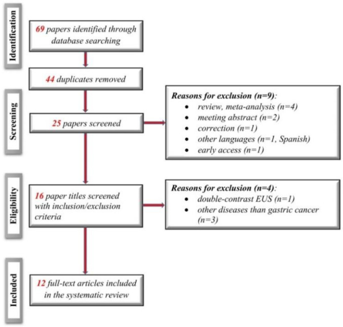
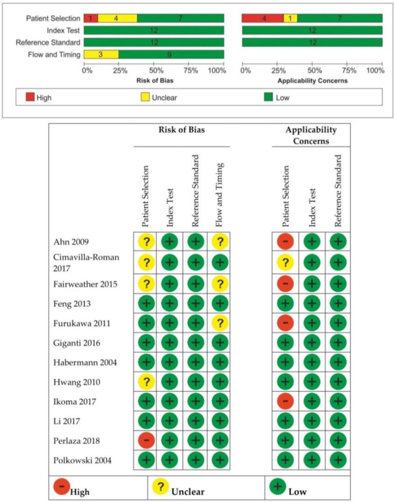
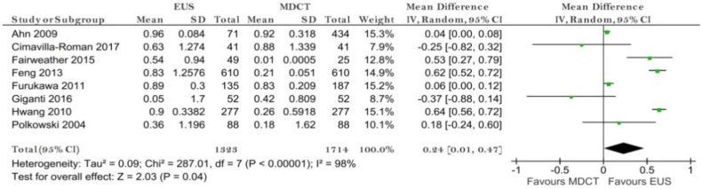
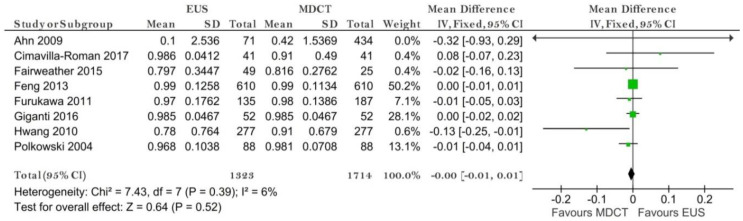
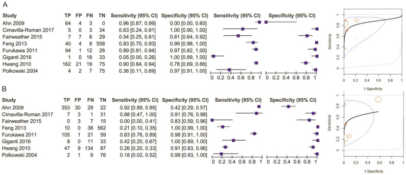
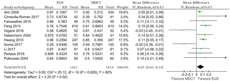
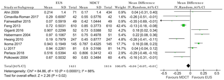
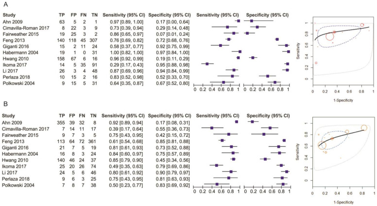
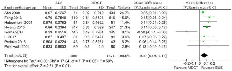
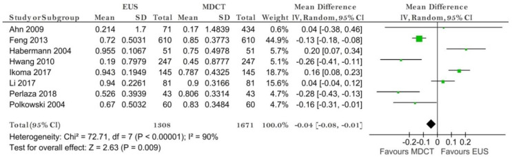
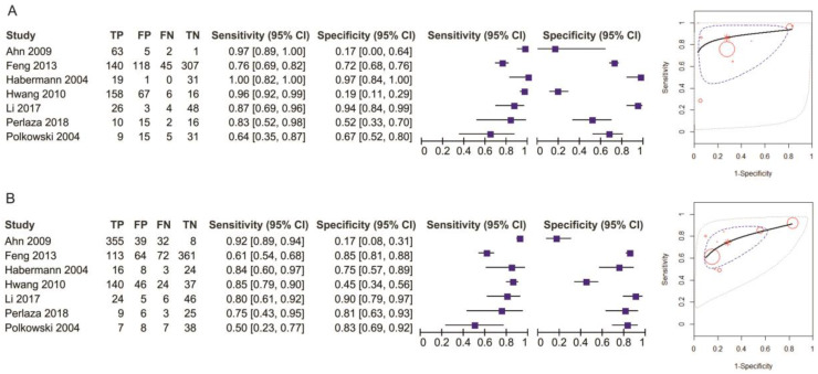
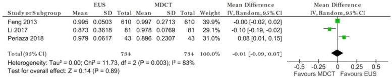
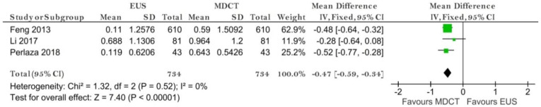
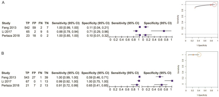
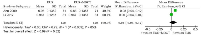
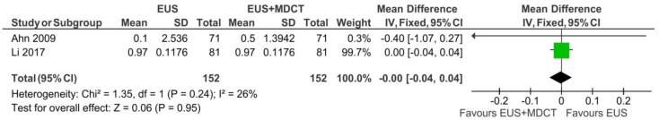
Gastric cancer preoperative staging is of outmost importance to assure proper management of the disease. Providing a relevant clinical stage relies on different imaging methods such as computed tomography (CT) or endoscopic ultrasound (EUS). We aimed to perform a network meta-analysis for gastric cancer clinical stage diagnostic tests, thus comparing the diagnostic accuracy of EUS vs. multidetector CT (MDCT) and EUS vs. EUS + MDCT. We plotted study estimates of pooled sensitivity and specificity on forest plots and summary receiver operating characteristic space to explore between-study variation in the performance of EUS, MDCT and EUS + MDCT for T1-T4, N0-N3, M0-M1 when data were available. Exploratory analyses were undertaken in RevMan 5. We included twelve studies with 2047 patients. Our results suggest that EUS was superior to MDCT in preoperative T1 and N staging. MDCT is more specific for the M stage but no significant difference in sensitivity was obtained. When comparing EUS vs. EUS + MDCT for T1 both sensitivity and specificity were not relevant. No significant differences were observed in T2-T4 stages. Even though EUS helped differentiate between the presence of invaded nodules, N stages should be carefully assessed by both methods since there is not sufficient data.

摘要

胃癌术前分期对于确保疾病的合理管理至关重要。提供相关的临床分期依赖于不同的成像方法,如计算机断层扫描(CT)或内镜超声(EUS)。我们旨在对胃癌临床分期诊断试验进行网状荟萃分析,从而比较EUS与多排CT(MDCT)以及EUS与EUS + MDCT的诊断准确性。当有数据时,我们在森林图和汇总接受者操作特征空间上绘制了合并敏感性和特异性的研究估计值,以探索EUS、MDCT和EUS + MDCT在T1 - T4、N0 - N3、M0 - M1分期表现中的研究间差异。在RevMan 5中进行了探索性分析。我们纳入了12项研究,共2047例患者。我们的结果表明,在术前T1和N分期方面,EUS优于MDCT。MDCT对M分期更具特异性,但在敏感性方面未获得显著差异。比较EUS与EUS + MDCT对T1分期时,敏感性和特异性均无相关性。在T2 - T4分期未观察到显著差异。尽管EUS有助于区分侵袭性结节的存在,但由于数据不足,两种方法都应仔细评估N分期。

https://cdn.ncbi.nlm.nih.gov/pmc/blobs/e4c4/7829791/993c50f5aac2/diagnostics-11-00134-g017.jpg
https://cdn.ncbi.nlm.nih.gov/pmc/blobs/e4c4/7829791/6646039dd403/diagnostics-11-00134-g001.jpg
https://cdn.ncbi.nlm.nih.gov/pmc/blobs/e4c4/7829791/0d359f58dde0/diagnostics-11-00134-g002.jpg
https://cdn.ncbi.nlm.nih.gov/pmc/blobs/e4c4/7829791/e9fba7202985/diagnostics-11-00134-g003.jpg
https://cdn.ncbi.nlm.nih.gov/pmc/blobs/e4c4/7829791/8db72bd1eb9f/diagnostics-11-00134-g004.jpg
https://cdn.ncbi.nlm.nih.gov/pmc/blobs/e4c4/7829791/9d822a994c55/diagnostics-11-00134-g005.jpg
https://cdn.ncbi.nlm.nih.gov/pmc/blobs/e4c4/7829791/abdf00a700bf/diagnostics-11-00134-g006.jpg
https://cdn.ncbi.nlm.nih.gov/pmc/blobs/e4c4/7829791/f174d7b37970/diagnostics-11-00134-g007.jpg
https://cdn.ncbi.nlm.nih.gov/pmc/blobs/e4c4/7829791/2c94c43668cb/diagnostics-11-00134-g008.jpg
https://cdn.ncbi.nlm.nih.gov/pmc/blobs/e4c4/7829791/6e927839f03f/diagnostics-11-00134-g009.jpg
https://cdn.ncbi.nlm.nih.gov/pmc/blobs/e4c4/7829791/3b180fdc391a/diagnostics-11-00134-g010.jpg
https://cdn.ncbi.nlm.nih.gov/pmc/blobs/e4c4/7829791/6b759422e5fb/diagnostics-11-00134-g011.jpg
https://cdn.ncbi.nlm.nih.gov/pmc/blobs/e4c4/7829791/f6ac79636ace/diagnostics-11-00134-g012.jpg
https://cdn.ncbi.nlm.nih.gov/pmc/blobs/e4c4/7829791/1bc0bc5bbb9f/diagnostics-11-00134-g013.jpg
https://cdn.ncbi.nlm.nih.gov/pmc/blobs/e4c4/7829791/5038fd2eb94c/diagnostics-11-00134-g014.jpg
https://cdn.ncbi.nlm.nih.gov/pmc/blobs/e4c4/7829791/94463dede569/diagnostics-11-00134-g015.jpg
https://cdn.ncbi.nlm.nih.gov/pmc/blobs/e4c4/7829791/844bb1d5f0ae/diagnostics-11-00134-g016.jpg
https://cdn.ncbi.nlm.nih.gov/pmc/blobs/e4c4/7829791/993c50f5aac2/diagnostics-11-00134-g017.jpg

相似文献

1
Endoscopic Ultrasound vs. Computed Tomography for Gastric Cancer Staging: A Network Meta-Analysis.内镜超声与计算机断层扫描用于胃癌分期:一项网状Meta分析
Diagnostics (Basel). 2021 Jan 16;11(1):134. doi: 10.3390/diagnostics11010134.
2
Endoscopic ultrasonography compared with multidetector computed tomography for the preoperative staging of gastric cancer: a meta-analysis.内镜超声检查与多排螺旋计算机断层扫描在胃癌术前分期中的比较:一项荟萃分析。
World J Surg Oncol. 2017 Jun 2;15(1):113. doi: 10.1186/s12957-017-1176-6.
3
Preoperative locoregional staging of gastric cancer: is there a place for magnetic resonance imaging? Prospective comparison with EUS and multidetector computed tomography.胃癌的术前局部区域分期:磁共振成像有一席之地吗?与超声内镜和多排螺旋计算机断层扫描的前瞻性比较
Gastric Cancer. 2016 Jan;19(1):216-25. doi: 10.1007/s10120-015-0468-1. Epub 2015 Jan 23.
4
Endoscopic ultrasound versus multidetector computed tomography in preoperative gastric cancer staging.内镜超声与多排螺旋计算机断层扫描在胃癌术前分期中的应用比较
Rev Esp Enferm Dig. 2017 Nov;109(11):761-767. doi: 10.17235/reed.2017.4638/2016.
5
Preoperative staging of gastric cancer by endoscopic ultrasonography and multidetector-row computed tomography.内镜超声检查和多排螺旋 CT 对胃癌的术前分期。
J Gastroenterol Hepatol. 2010 Mar;25(3):512-8. doi: 10.1111/j.1440-1746.2009.06106.x.
6
Prospective comparison of MR with diffusion-weighted imaging, endoscopic ultrasound, MDCT and positron emission tomography-CT in the pre-operative staging of oesophageal cancer: results from a pilot study.磁共振成像与扩散加权成像、内镜超声、多层螺旋CT及正电子发射断层扫描-CT在食管癌术前分期中的前瞻性比较:一项初步研究结果
Br J Radiol. 2016 Dec;89(1068):20160087. doi: 10.1259/bjr.20160087. Epub 2016 Oct 21.
7
Staging of rectal carcinoma: MDCT, MRI or EUS. Single center experience.直肠癌的分期:多层螺旋CT、磁共振成像或超声内镜检查。单中心经验。
Turk J Gastroenterol. 2014 Dec;25(6):669-73. doi: 10.5152/tjg.2014.6214.
8
Imaging in local staging of gastric cancer: a systematic review.胃癌局部分期的影像学检查:一项系统评价
J Clin Oncol. 2007 May 20;25(15):2107-16. doi: 10.1200/JCO.2006.09.5224.
9
Preoperative Tumor Staging of Gastric Cancer: Comparison of Double Contrast-Enhanced Ultrasound and Multidetector Computed Tomography.胃癌术前肿瘤分期:双重对比增强超声与多排螺旋 CT 的比较。
J Ultrasound Med. 2019 Dec;38(12):3203-3209. doi: 10.1002/jum.15028. Epub 2019 May 21.
10
Gastric cancer-can we rely only on endoscopic ultrasonography?胃癌——我们能否仅依靠内镜超声检查?
Endosc Ultrasound. 2014 Apr;3(Suppl 1):S3.

引用本文的文献

1
Endoscopic Ultrasound (EUS) in Gastric Cancer: Current Applications and Future Perspectives.胃癌中的内镜超声(EUS):当前应用与未来展望
Diseases. 2025 Jul 24;13(8):234. doi: 10.3390/diseases13080234.
2
Serum metabolic profiling analysis of chronic gastritis and gastric cancer by untargeted metabolomics.基于非靶向代谢组学的慢性胃炎和胃癌血清代谢谱分析
Ann Med Surg (Lond). 2024 Dec 19;87(2):583-597. doi: 10.1097/MS9.0000000000002977. eCollection 2025 Feb.
3
Nomogram for pre-procedural prediction of lymph node metastasis in patients with submucosal early gastric cancer.

本文引用的文献

1
Gastric cancer.胃癌。
Lancet. 2020 Aug 29;396(10251):635-648. doi: 10.1016/S0140-6736(20)31288-5.
2
Quantification of Structural Heterogeneity Using Fractal Analysis of Contrast-Enhanced CT Image to Predict Survival in Gastric Cancer Patients.使用增强 CT 图像分形分析量化结构异质性预测胃癌患者生存。
Dig Dis Sci. 2021 Jun;66(6):2069-2074. doi: 10.1007/s10620-020-06479-w. Epub 2020 Jul 20.
3
Preoperative computed tomography-guided disease-free survival prediction in gastric cancer: a multicenter radiomics study.
黏膜下早期胃癌患者术前淋巴结转移预测列线图
Surg Endosc. 2025 Mar;39(3):1661-1671. doi: 10.1007/s00464-024-11517-z. Epub 2025 Jan 9.
4
Diagnostic Efficacy of Staging Laparoscopy Compared to CT and PET-CT in Gastric Cancer: A Retrospective Cohort Analysis.分期腹腔镜检查与CT及PET-CT在胃癌诊断中的效能比较:一项回顾性队列分析
Medicina (Kaunas). 2024 Dec 19;60(12):2079. doi: 10.3390/medicina60122079.
5
Influence of visceral adipose tissue on the accuracy of tumor T-staging of gastric cancer in preoperative CT.内脏脂肪组织对术前CT胃癌肿瘤T分期准确性的影响。
Jpn J Radiol. 2025 Apr;43(4):656-665. doi: 10.1007/s11604-024-01711-3. Epub 2024 Nov 28.
6
Double contrast-enhanced ultrasonography improves diagnostic accuracy of T staging compared with multi-detector computed tomography in gastric cancer patients.双重对比增强超声检查提高了胃癌患者 T 分期的诊断准确性,优于多排螺旋 CT。
World J Gastroenterol. 2024 Jun 21;30(23):3005-3015. doi: 10.3748/wjg.v30.i23.3005.
7
Development of a deep learning model for early gastric cancer diagnosis using preoperative computed tomography images.使用术前计算机断层扫描图像开发用于早期胃癌诊断的深度学习模型。
Front Oncol. 2023 Oct 6;13:1265366. doi: 10.3389/fonc.2023.1265366. eCollection 2023.
8
Linear EUS Accuracy in Preoperative Staging of Gastric Cancer: A Retrospective Multicenter Study.线性超声内镜在胃癌术前分期中的准确性:一项回顾性多中心研究
Diagnostics (Basel). 2023 May 25;13(11):1842. doi: 10.3390/diagnostics13111842.
9
Imaging in Gastric Cancer: Current Practice and Future Perspectives.胃癌的影像学检查:当前实践与未来展望
Diagnostics (Basel). 2023 Mar 28;13(7):1276. doi: 10.3390/diagnostics13071276.
10
Detection and Characterization of Gastric Cancer Using Cascade Deep Learning Model in Endoscopic Images.使用级联深度学习模型在内窥镜图像中检测和表征胃癌
Diagnostics (Basel). 2022 Aug 18;12(8):1996. doi: 10.3390/diagnostics12081996.
术前计算机断层扫描引导的胃癌无病生存预测:一项多中心放射组学研究
Med Phys. 2020 Oct;47(10):4862-4871. doi: 10.1002/mp.14350. Epub 2020 Aug 5.
4
Hierarchical Analysis of Factors Associated with T Staging of Gastric Cancer by Endoscopic Ultrasound.基于内镜超声的胃癌 T 分期相关因素的层次分析。
Dig Dis Sci. 2021 Feb;66(2):612-618. doi: 10.1007/s10620-020-06194-6. Epub 2020 Mar 17.
5
A Meta-Analysis And Systematic Review Of Accuracy Of Endoscopic Ultrasound For N Staging Of Gastric Cancers.一项关于超声内镜对胃癌N分期准确性的Meta分析与系统评价
Cancer Manag Res. 2019 Sep 27;11:8755-8764. doi: 10.2147/CMAR.S200318. eCollection 2019.
6
Evidence Based Surgical Approach to Locally Advanced Gastric Cancer.局部进展期胃癌的循证外科治疗方法
J Nepal Health Res Counc. 2019 Aug 4;17(2):133-140. doi: 10.33314/jnhrc.v0i0.2055.
7
Linear-array endoscopic ultrasound improves the accuracy of preoperative submucosal invasion prediction in suspected early gastric cancer compared with radial endoscopic ultrasound: A prospective cohort study.线性内镜超声提高了疑似早期胃癌术前黏膜下浸润预测的准确性,优于径向内镜超声:一项前瞻性队列研究。
J Gastroenterol Hepatol. 2020 Jan;35(1):118-123. doi: 10.1111/jgh.14819. Epub 2019 Sep 1.
8
Gastric cancer and imaging biomarkers: Part 1 - a critical review of DW-MRI and CE-MDCT findings.胃癌与影像学标志物:第一部分——弥散加权 MRI 和 CE-MDCT 研究结果的批判性综述。
Eur Radiol. 2019 Apr;29(4):1743-1753. doi: 10.1007/s00330-018-5732-4. Epub 2018 Oct 2.
9
Should 18F-FDG PET/CT Be Routinely Performed in the Clinical Staging of Locally Advanced Gastric Adenocarcinoma?18F-FDG PET/CT 在局部进展期胃腺癌临床分期中是否应常规进行?
Clin Nucl Med. 2018 Jun;43(6):402-410. doi: 10.1097/RLU.0000000000002028.
10
Clinical efficacy of endoscopic ultrasonography for decision of treatment strategy of gastric cancer.内镜超声检查对胃癌治疗策略决策的临床疗效。
Surg Endosc. 2018 Sep;32(9):3789-3797. doi: 10.1007/s00464-018-6104-5. Epub 2018 Feb 12.