Feng Kang, Zhang Yu, Chen Yue-Guo
Department of Ophthalmology, Peking University Third Hospital, 49 North Garden Road, Haidian District, Beijing, 100191, People's Republic of China.
Beijing Key Laboratory of Restoration of Damaged Ocular Nerve, Beijing, China.
BMC Ophthalmol. 2021 Jan 19;21(1):47. doi: 10.1186/s12886-021-01806-9.
To explore the possible causes of tomography suspect keratoconus (TSK) marked by Tomography in screening keratoconus in a Chinese cohort, and the reasonable range of corneal horizontal diameter and thickness for decreasing the proportion of TSK.
Nested case-control study from a single center prospective cohort. All subjects were selected from the Peking University Third Hospital Ectasia Cornea Disease Cohort Project database, which included myopic patients seeking corneal refractive surgical corrections since 2013. Demographic information, basic eye examination, and auxiliary equipment examination including refraction, IOL-master, Pentacam, Sirius, and Topolyzer were recorded. In this study, all cases were classified into two groups: TSK group and normal control (NC) group, and all of them were followed up at least 2 years. The former is consisted of those whose screening examinations of tomography are abnormal, the latter is those whose screening examinations are normal. All of them have already been followed up at least 2 years without abnormalities after excimer laser corneal refractive surgeries. Unpaired t tests and Chi-square tests were used to compare the differences of indices from the tomography between the two groups.
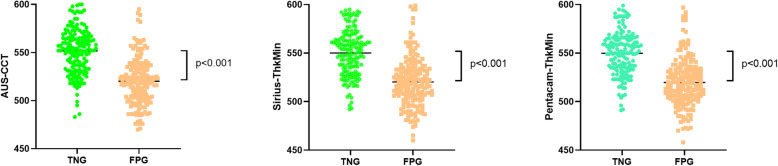
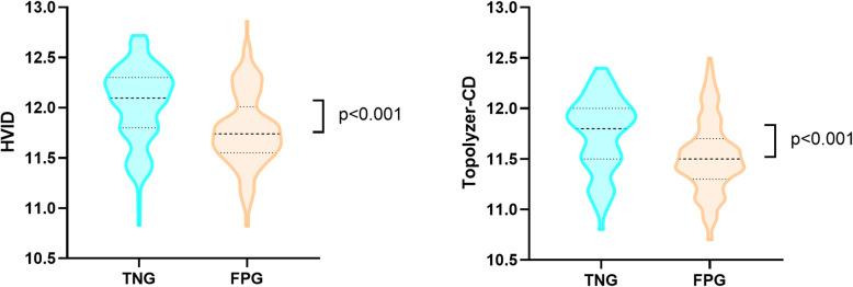
Of 183 TSK eyes (109 patients) and 160 NC eyes (83 patients), the mean age is 28.0 and 26.3 years old respectively. The corneal horizontal diameter is 11.5-11.8 mm in TSK group and 11.8-12.0 mm in NC group. The central corneal thickness is nearly 520 μm in the former and 550 μm in the latter. For Sirius, the TSK ratio of indices of SIf and SIb is 41.5 and 39.9% respectively in TSK group. For Pentacam, the TSK ratio of index IHD is 59.0% and "final D" is 72.7%.
Corneal horizontal diameter and central corneal thickness have great influences on the results of corneal tomography in detecting the suspect keratoconus.
在中国人群中,探讨在圆锥角膜筛查中,以断层扫描标记的断层扫描可疑圆锥角膜(TSK)的可能病因,以及降低TSK比例的角膜水平直径和厚度的合理范围。
来自单中心前瞻性队列的巢式病例对照研究。所有受试者均选自北京大学第三医院角膜扩张疾病队列项目数据库,该数据库包括自2013年以来寻求角膜屈光手术矫正的近视患者。记录人口统计学信息、基本眼部检查以及包括验光、IOL-master、Pentacam、Sirius和Topolyzer在内的辅助设备检查结果。在本研究中,所有病例分为两组:TSK组和正常对照组(NC组),两组均至少随访2年。前者由断层扫描筛查检查异常者组成,后者由筛查检查正常者组成。所有患者在准分子激光角膜屈光手术后至少随访2年且无异常。采用独立样本t检验和卡方检验比较两组断层扫描指标的差异。
在183只TSK眼(109例患者)和160只NC眼(83例患者)中,平均年龄分别为28.0岁和26.3岁。TSK组角膜水平直径为11.5 - 11.8mm,NC组为11.8 - 12.0mm。前者中央角膜厚度近520μm,后者为550μm。对于Sirius,TSK组SIf和SIb指标的TSK比例分别为41.5%和39.9%。对于Pentacam,指标IHD的TSK比例为59.0%,“最终D”为72.7%。
角膜水平直径和中央角膜厚度对角膜断层扫描检测可疑圆锥角膜的结果有很大影响。