Geden Matthew J, Romero Selena E, Deshmukh Mohanish
Department of Cell Biology and Physiology, University of North Carolina, Chapel Hill, NC, 27599, USA.
Neuroscience Center, University of North Carolina, Chapel Hill, NC, 27599, USA.
Cell Death Dis. 2021 Jan 20;12(1):104. doi: 10.1038/s41419-020-03373-1.
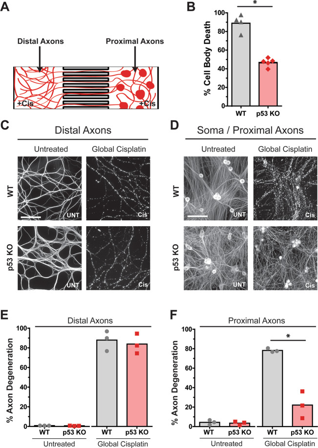
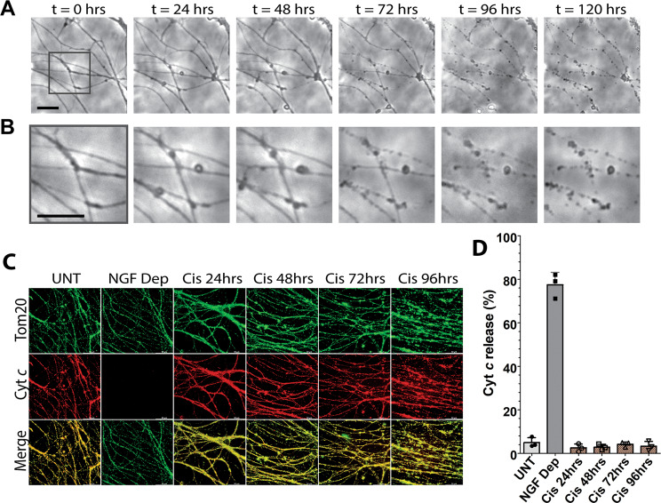
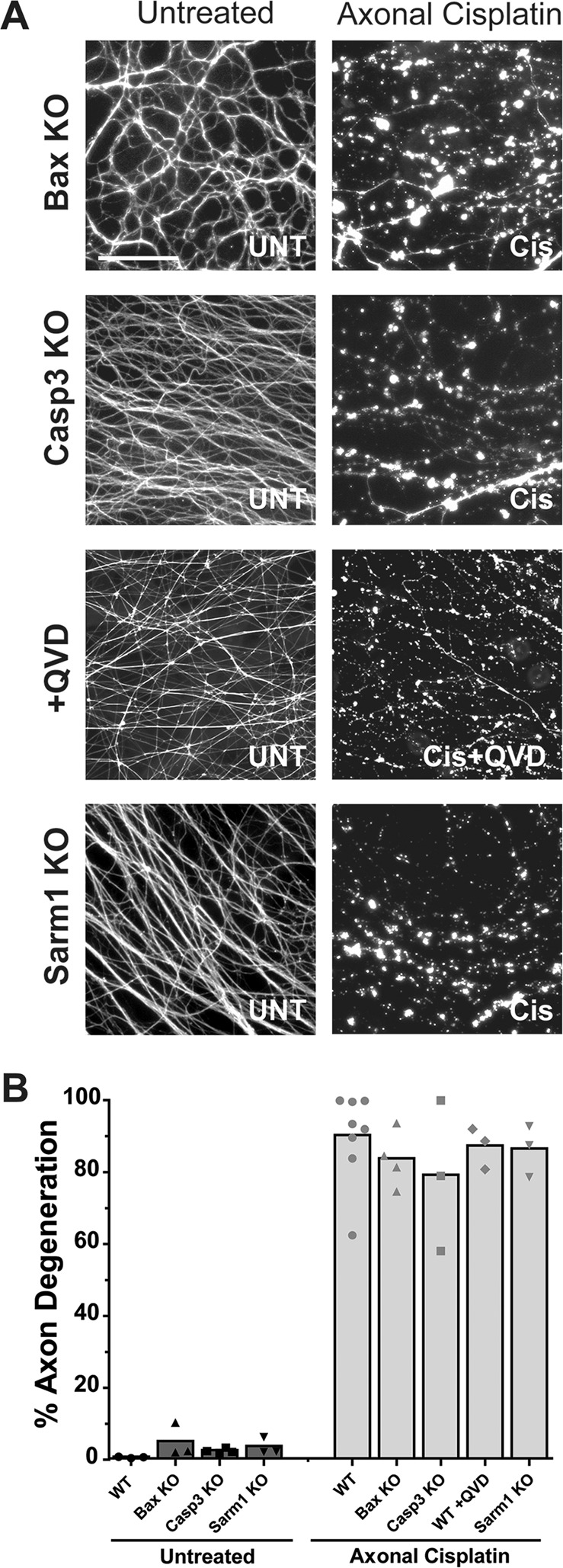
While the consequences of nuclear DNA damage have been well studied, the exact consequences of acute and selective mitochondrial DNA (mtDNA) damage are less understood. DNA damaging chemotherapeutic drugs are known to activate p53-dependent apoptosis in response to sustained nuclear DNA damage. While it is recognized that whole-cell exposure to these drugs also damages mtDNA, the specific contribution of mtDNA damage to cellular degeneration is less clear. To examine this, we induced selective mtDNA damage in neuronal axons using microfluidic chambers that allow for the spatial and fluidic isolation of neuronal cell bodies (containing nucleus and mitochondria) from the axons (containing mitochondria). Exposure of the DNA damaging drug cisplatin selectively to only the axons induced mtDNA damage in axonal mitochondria, without nuclear damage. We found that this resulted in the selective degeneration of only the targeted axons that were exposed to DNA damage, where ROS was induced but mitochondria were not permeabilized. mtDNA damage-induced axon degeneration was not mediated by any of the three known axon degeneration pathways: apoptosis, axon pruning, and Wallerian degeneration, as Bax-deficiency, or Casp3-deficiency, or Sarm1-deficiency failed to protect the degenerating axons. Strikingly, p53, which is essential for degeneration after nuclear DNA damage, was also not required for degeneration induced with mtDNA damage. This was most evident when the p53-deficient neurons were globally exposed to cisplatin. While the cell bodies of p53-deficient neurons were protected from degeneration in this context, the axons farthest from the cell bodies still underwent degeneration. These results highlight how whole cell exposure to DNA damage activates two pathways of degeneration; a faster, p53-dependent apoptotic degeneration that is triggered in the cell bodies with nuclear DNA damage, and a slower, p53-independent degeneration that is induced with mtDNA damage.
虽然核DNA损伤的后果已得到充分研究,但急性和选择性线粒体DNA(mtDNA)损伤的确切后果却鲜为人知。已知DNA损伤化疗药物会在持续的核DNA损伤时激活p53依赖性凋亡。虽然人们认识到全细胞暴露于这些药物也会损伤mtDNA,但mtDNA损伤对细胞退化的具体作用尚不清楚。为了研究这一点,我们使用微流控室在神经元轴突中诱导选择性mtDNA损伤,该微流控室允许将神经元细胞体(包含细胞核和线粒体)与轴突(包含线粒体)进行空间和流体隔离。将DNA损伤药物顺铂仅选择性地暴露于轴突会诱导轴突线粒体中的mtDNA损伤,而不会造成核损伤。我们发现,这仅导致暴露于DNA损伤的靶向轴突发生选择性退化,其中会诱导活性氧(ROS)产生,但线粒体未发生通透性改变。mtDNA损伤诱导的轴突退化并非由三种已知的轴突退化途径中的任何一种介导:凋亡、轴突修剪和沃勒变性,因为Bax缺陷、Casp3缺陷或Sarm1缺陷均无法保护退化的轴突。令人惊讶的是,对于核DNA损伤后的退化至关重要的p53,对于mtDNA损伤诱导的退化也不是必需的。当p53缺陷的神经元整体暴露于顺铂时,这一点最为明显。在此情况下,p53缺陷神经元的细胞体免受退化,但离细胞体最远的轴突仍会发生退化。这些结果突出了全细胞暴露于DNA损伤如何激活两种退化途径;一种更快的、p53依赖性凋亡性退化,由细胞核DNA损伤在细胞体中触发,以及一种较慢的、p53非依赖性退化,由mtDNA损伤诱导。