• 文献检索
  • 文档翻译
  • 深度研究
  • 学术资讯
  • Suppr Zotero 插件Zotero 插件
  • 邀请有礼
  • 套餐&价格
  • 历史记录
应用&插件
Suppr Zotero 插件Zotero 插件浏览器插件Mac 客户端Windows 客户端微信小程序
定价
高级版会员购买积分包购买API积分包
服务
文献检索文档翻译深度研究API 文档MCP 服务
关于我们
关于 Suppr公司介绍联系我们用户协议隐私条款
关注我们

Suppr 超能文献

核心技术专利:CN118964589B侵权必究
粤ICP备2023148730 号-1Suppr @ 2026

文献检索

告别复杂PubMed语法,用中文像聊天一样搜索,搜遍4000万医学文献。AI智能推荐,让科研检索更轻松。

立即免费搜索

文件翻译

保留排版,准确专业,支持PDF/Word/PPT等文件格式,支持 12+语言互译。

免费翻译文档

深度研究

AI帮你快速写综述,25分钟生成高质量综述,智能提取关键信息,辅助科研写作。

立即免费体验

壳聚糖与嗅黏膜间充质干/基质细胞联合应用促进周围神经再生

Combined Use of Chitosan and Olfactory Mucosa Mesenchymal Stem/Stromal Cells to Promote Peripheral Nerve Regeneration .

作者信息

Alvites Rui D, Branquinho Mariana V, Sousa Ana C, Amorim Irina, Magalhães Rui, João Filipa, Almeida Diogo, Amado Sandra, Prada Justina, Pires Isabel, Zen Federica, Raimondo Stefania, Luís Ana L, Geuna Stefano, Varejão Artur S P, Maurício Ana C

机构信息

Departamento de Clínicas Veterinárias, Instituto de Ciências Biomédicas de Abel Salazar (ICBAS), Universidade do Porto (UP), Rua de Jorge Viterbo Ferreira, No. 228, 4050-313 Porto, Portugal.

Centro de Estudos de Ciência Animal (CECA), Instituto de Ciências, Tecnologias e Agroambiente da Universidade do Porto (ICETA), Rua D. Manuel II, Apartado 55142, 4051-401 Porto, Portugal.

出版信息

Stem Cells Int. 2021 Jan 2;2021:6613029. doi: 10.1155/2021/6613029. eCollection 2021.

DOI:10.1155/2021/6613029
PMID:33488738
原文链接:https://pmc.ncbi.nlm.nih.gov/articles/PMC7801080/
Abstract

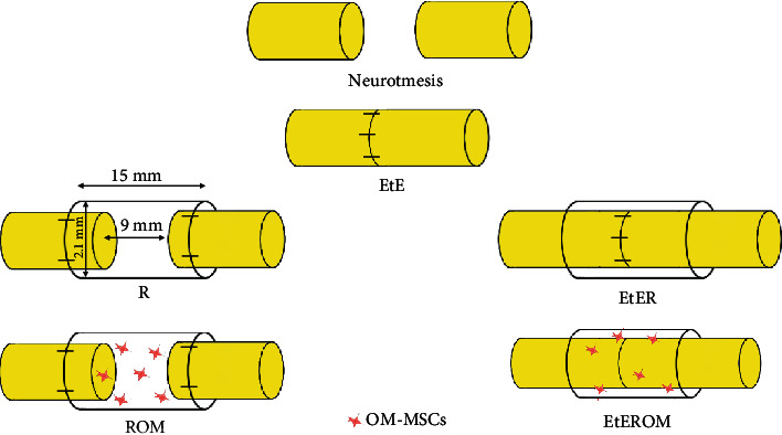
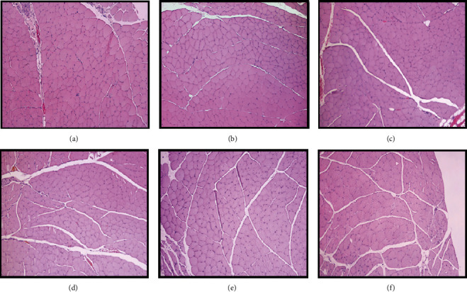
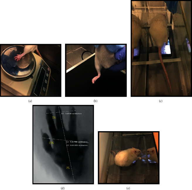
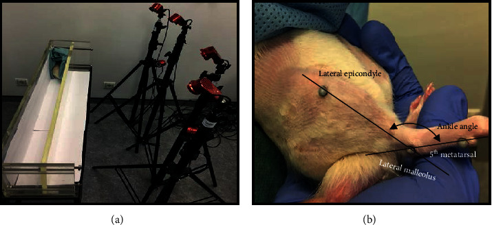
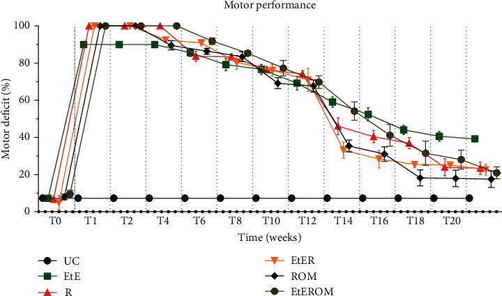
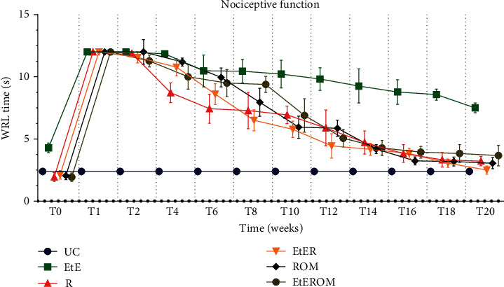
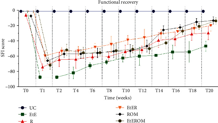
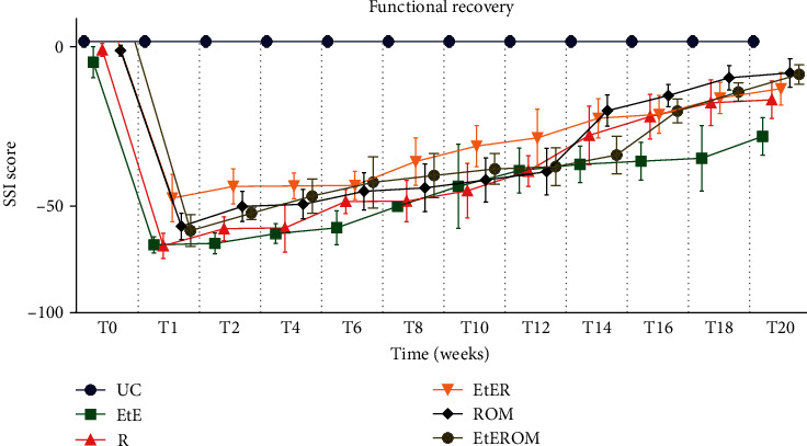
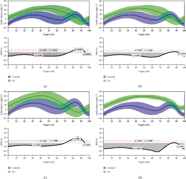
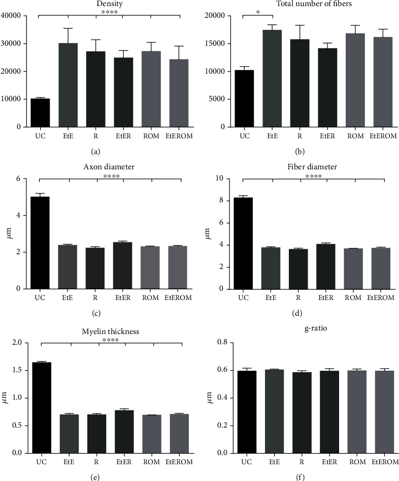
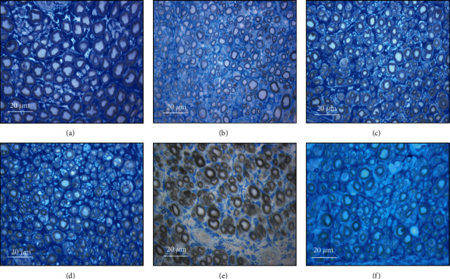
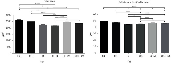
Peripheral nerve injury remains a clinical challenge with severe physiological and functional consequences. Despite the existence of multiple possible therapeutic approaches, until now, there is no consensus regarding the advantages of each option or the best methodology in promoting nerve regeneration. Regenerative medicine is a promise to overcome this medical limitation, and in this work, chitosan nerve guide conduits and olfactory mucosa mesenchymal stem/stromal cells were applied in different therapeutic combinations to promote regeneration in sciatic nerves after neurotmesis injury. Over 20 weeks, the intervened animals were subjected to a regular functional assessment (determination of motor performance, nociception, and sciatic indexes), and after this period, they were evaluated kinematically and the sciatic nerves and cranial tibial muscles were evaluated stereologically and histomorphometrically, respectively. The results obtained allowed confirming the beneficial effects of using these therapeutic approaches. The use of chitosan NGCs and cells resulted in better motor performance, better sciatic indexes, and lower gait dysfunction after 20 weeks. The use of only NGGs demonstrated better nociceptive recoveries. The stereological evaluation of the sciatic nerve revealed identical values in the different parameters for all therapeutic groups. In the muscle histomorphometric evaluation, the groups treated with NGCs and cells showed results close to those of the group that received traditional sutures, the one with the best final values. The therapeutic combinations studied show promising outcomes and should be the target of new future works to overcome some irregularities found in the results and establish the combination of nerve guidance conduits and olfactory mucosa mesenchymal stem/stromal cells as viable options in the treatment of peripheral nerves after injury.

摘要

周围神经损伤仍然是一个临床挑战,会带来严重的生理和功能后果。尽管存在多种可能的治疗方法,但到目前为止,对于每种选择的优势或促进神经再生的最佳方法尚未达成共识。再生医学有望克服这一医学局限,在本研究中,壳聚糖神经导管和嗅黏膜间充质干/基质细胞被应用于不同的治疗组合,以促进坐骨神经切断伤后的再生。在20周的时间里,对干预后的动物进行定期功能评估(测定运动性能、痛觉感受和坐骨神经指数),在此期间过后,对它们进行运动学评估,并分别对坐骨神经和胫前肌进行体视学和组织形态计量学评估。所获得的结果证实了使用这些治疗方法的有益效果。使用壳聚糖神经导管和细胞在20周后带来了更好的运动性能、更好的坐骨神经指数和更低的步态功能障碍。仅使用神经导管显示出更好的痛觉恢复。坐骨神经的体视学评估显示,所有治疗组在不同参数上的值相同。在肌肉组织形态计量学评估中,用神经导管和细胞治疗的组显示出的结果与接受传统缝合的组相近,而传统缝合组的最终值最佳。所研究的治疗组合显示出有前景的结果,应该成为未来新研究的目标,以克服结果中发现的一些不规则之处,并将神经引导导管和嗅黏膜间充质干/基质细胞的组合确立为损伤后周围神经治疗的可行选择。

https://cdn.ncbi.nlm.nih.gov/pmc/blobs/e408/7801080/a9401a27c5d1/SCI2021-6613029.018.jpg
https://cdn.ncbi.nlm.nih.gov/pmc/blobs/e408/7801080/4d7daf5dfe4a/SCI2021-6613029.001.jpg
https://cdn.ncbi.nlm.nih.gov/pmc/blobs/e408/7801080/8b82f055cbb4/SCI2021-6613029.002.jpg
https://cdn.ncbi.nlm.nih.gov/pmc/blobs/e408/7801080/d040a740a570/SCI2021-6613029.003.jpg
https://cdn.ncbi.nlm.nih.gov/pmc/blobs/e408/7801080/88b054a16c6b/SCI2021-6613029.004.jpg
https://cdn.ncbi.nlm.nih.gov/pmc/blobs/e408/7801080/616bfcb24549/SCI2021-6613029.005.jpg
https://cdn.ncbi.nlm.nih.gov/pmc/blobs/e408/7801080/038cba010560/SCI2021-6613029.006.jpg
https://cdn.ncbi.nlm.nih.gov/pmc/blobs/e408/7801080/e2bcf14249d8/SCI2021-6613029.007.jpg
https://cdn.ncbi.nlm.nih.gov/pmc/blobs/e408/7801080/b3134be5d576/SCI2021-6613029.008.jpg
https://cdn.ncbi.nlm.nih.gov/pmc/blobs/e408/7801080/2ff4b6985839/SCI2021-6613029.009.jpg
https://cdn.ncbi.nlm.nih.gov/pmc/blobs/e408/7801080/f4624c4329c1/SCI2021-6613029.010.jpg
https://cdn.ncbi.nlm.nih.gov/pmc/blobs/e408/7801080/414e235bf566/SCI2021-6613029.011.jpg
https://cdn.ncbi.nlm.nih.gov/pmc/blobs/e408/7801080/08994143ec79/SCI2021-6613029.012.jpg
https://cdn.ncbi.nlm.nih.gov/pmc/blobs/e408/7801080/7a59b1265b94/SCI2021-6613029.013.jpg
https://cdn.ncbi.nlm.nih.gov/pmc/blobs/e408/7801080/828fcced5a53/SCI2021-6613029.014.jpg
https://cdn.ncbi.nlm.nih.gov/pmc/blobs/e408/7801080/429b4b9773a8/SCI2021-6613029.015.jpg
https://cdn.ncbi.nlm.nih.gov/pmc/blobs/e408/7801080/d3c31e21abc8/SCI2021-6613029.016.jpg
https://cdn.ncbi.nlm.nih.gov/pmc/blobs/e408/7801080/586143bae7d2/SCI2021-6613029.017.jpg
https://cdn.ncbi.nlm.nih.gov/pmc/blobs/e408/7801080/a9401a27c5d1/SCI2021-6613029.018.jpg

相似文献

1
Combined Use of Chitosan and Olfactory Mucosa Mesenchymal Stem/Stromal Cells to Promote Peripheral Nerve Regeneration .壳聚糖与嗅黏膜间充质干/基质细胞联合应用促进周围神经再生
Stem Cells Int. 2021 Jan 2;2021:6613029. doi: 10.1155/2021/6613029. eCollection 2021.
2
Effects of Olfactory Mucosa Stem/Stromal Cell and Olfactory Ensheating Cells Secretome on Peripheral Nerve Regeneration.嗅黏膜干/基质细胞和嗅鞘细胞分泌组对周围神经再生的影响
Biomolecules. 2022 Jun 11;12(6):818. doi: 10.3390/biom12060818.
3
Nanofibrous Nerve Conduits with Nerve Growth Factors and Bone Marrow Stromal Cells Pre-Cultured in Bioreactors for Peripheral Nerve Regeneration.生物反应器中预培养神经生长因子和骨髓基质细胞的纳米纤维神经导管用于周围神经再生。
ACS Appl Mater Interfaces. 2020 Apr 8;12(14):16168-16177. doi: 10.1021/acsami.0c04191. Epub 2020 Mar 25.
4
Use of hybrid chitosan membranes and human mesenchymal stem cells from the Wharton jelly of umbilical cord for promoting nerve regeneration in an axonotmesis rat model.使用杂交壳聚糖膜和脐带华通氏胶来源的人间质干细胞促进轴索切断大鼠模型中的神经再生。
Neural Regen Res. 2012 Oct 15;7(29):2247-58. doi: 10.3969/j.issn.1673-5374.2012.29.002.
5
Chitosan-film associated with mesenchymal stem cells enhanced regeneration of peripheral nerves: A rat sciatic nerve model.壳聚糖膜联合间充质干细胞促进周围神经再生:大鼠坐骨神经模型
J Chem Neuroanat. 2018 Mar;88:46-54. doi: 10.1016/j.jchemneu.2017.10.003. Epub 2017 Oct 26.
6
Regenerative potential of chitosan-coated poly-3-hydroxybutyrate conduits seeded with mesenchymal stem cells in a rat sciatic nerve injury model.壳聚糖包被的聚-3-羟基丁酸酯导管接种间充质干细胞在大鼠坐骨神经损伤模型中的再生潜力
Int J Neurosci. 2018 Sep;128(9):828-834. doi: 10.1080/00207454.2018.1435536. Epub 2018 Feb 15.
7
Peripheral nerve repair: monitoring by using gadofluorine M-enhanced MR imaging with chitosan nerve conduits with cultured mesenchymal stem cells in rat model of neurotmesis.周围神经修复:壳聚糖神经导管联合培养的间充质干细胞在大鼠神经横断模型中通过钆弗塞胺增强 MRI 监测。
Radiology. 2012 Jan;262(1):161-71. doi: 10.1148/radiol.11110911. Epub 2011 Nov 4.
8
Combined Wharton's jelly derived mesenchymal stem cells and nerve guidance conduit: A potential promising therapy for peripheral nerve injuries.联合沃顿胶源性间充质干细胞与神经导管:一种治疗周围神经损伤的潜在有效疗法。
Int J Biochem Cell Biol. 2017 May;86:67-76. doi: 10.1016/j.biocel.2017.03.002. Epub 2017 Mar 6.
9
Schwann cells and mesenchymal stem cells in laminin- or fibronectin-aligned matrices and regeneration across a critical size defect of 15 mm in the rat sciatic nerve.层粘连蛋白或纤连蛋白排列基质中的雪旺细胞和间充质干细胞与大鼠坐骨神经15毫米临界尺寸缺损处的再生
J Neurosurg Spine. 2018 Jan;28(1):109-118. doi: 10.3171/2017.5.SPINE161100. Epub 2017 Nov 10.
10
Functional collagen conduits combined with human mesenchymal stem cells promote regeneration after sciatic nerve transection in dogs.功能胶原导管联合人骨髓间充质干细胞促进犬坐骨神经横断后再生。
J Tissue Eng Regen Med. 2018 May;12(5):1285-1296. doi: 10.1002/term.2660. Epub 2018 Mar 25.

引用本文的文献

1
Mesenchymal stem cell-based therapy for peripheral nerve injuries: A promise or reality?基于间充质干细胞的外周神经损伤治疗:是希望还是现实?
World J Stem Cells. 2025 Jun 26;17(6):107833. doi: 10.4252/wjsc.v17.i6.107833.
2
Combined use of olfactory mucosal mesenchymal stem cells conditioned medium and neural guide conduits promotes nerve regeneration in an ovine model.嗅黏膜间充质干细胞条件培养基与神经引导导管联合应用促进绵羊模型中的神经再生。
Front Cell Dev Biol. 2025 May 2;13:1598736. doi: 10.3389/fcell.2025.1598736. eCollection 2025.
3
Biomechanical Evaluation of the Sheep Common Peroneal Nerve After Crush Injury.

本文引用的文献

1
Rat Olfactory Mucosa Mesenchymal Stem/Stromal Cells (OM-MSCs): A Characterization Study.大鼠嗅黏膜间充质干/基质细胞(OM-MSCs):一项特性研究。
Int J Cell Biol. 2020 Jan 29;2020:2938258. doi: 10.1155/2020/2938258. eCollection 2020.
2
Restoration of Neurological Function Following Peripheral Nerve Trauma.周围神经损伤后的神经功能恢复。
Int J Mol Sci. 2020 Mar 6;21(5):1808. doi: 10.3390/ijms21051808.
3
Multi-target approaches to CNS repair: olfactory mucosa-derived cells and heparan sulfates.中枢神经系统修复的多靶点方法:嗅黏膜来源的细胞和硫酸乙酰肝素。
挤压伤后绵羊腓总神经的生物力学评估
Animals (Basel). 2025 Feb 21;15(5):627. doi: 10.3390/ani15050627.
4
evaluation of dental pulp stem cells for sciatic nerve regeneration: foundations for future applications.用于坐骨神经再生的牙髓干细胞评估:未来应用的基础
Front Cell Dev Biol. 2025 Feb 19;13:1528213. doi: 10.3389/fcell.2025.1528213. eCollection 2025.
5
Marine bioactives: a new frontier in neurosurgical therapeutics.海洋生物活性物质:神经外科学治疗的新领域。
Neurosurg Rev. 2024 Oct 12;47(1):790. doi: 10.1007/s10143-024-03034-3.
6
Development of Biomaterials for Addressing Upper Extremity Peripheral Nerve Gaps.用于解决上肢周围神经缺损的生物材料的研发
J Hand Surg Glob Online. 2024 Mar 27;6(5):711-717. doi: 10.1016/j.jhsg.2024.01.023. eCollection 2024 Sep.
7
Recent perspectives on the synergy of mesenchymal stem cells with micro/nano strategies in peripheral nerve regeneration-a review.间充质干细胞与微/纳米策略在外周神经再生中的协同作用研究新进展——综述
Front Bioeng Biotechnol. 2024 Jul 10;12:1401512. doi: 10.3389/fbioe.2024.1401512. eCollection 2024.
8
Mechanism and treatment of olfactory dysfunction caused by coronavirus disease 2019.新型冠状病毒病导致嗅觉功能障碍的机制和治疗。
J Transl Med. 2023 Nov 17;21(1):829. doi: 10.1186/s12967-023-04719-x.
9
Ultrasound Landmarks in the Approach to the Common Peroneal Nerve in a Sheep Model-Application in Peripheral Nerve Regeneration.绵羊模型中腓总神经入路的超声标志——在外周神经再生中的应用
Life (Basel). 2023 Sep 15;13(9):1919. doi: 10.3390/life13091919.
10
Whole body vibration accelerates the functional recovery of motor nerve components in sciatic nerve-crush injury model rats.全身振动可加速坐骨神经挤压伤模型大鼠运动神经成分的功能恢复。
J Exerc Rehabil. 2023 Jun 28;19(3):149-162. doi: 10.12965/jer.2346178.089. eCollection 2023 Jun.
Nat Rev Neurol. 2020 Apr;16(4):229-240. doi: 10.1038/s41582-020-0311-0. Epub 2020 Feb 25.
4
M1 macrophage infiltration exacerbate muscle/bone atrophy after peripheral nerve injury.M1 巨噬细胞浸润加剧外周神经损伤后的肌肉/骨萎缩。
BMC Musculoskelet Disord. 2020 Jan 20;21(1):44. doi: 10.1186/s12891-020-3069-z.
5
Dextran-based tube-guides for the regeneration of the rat sciatic nerve after neurotmesis injury.基于葡聚糖的管引导物在大鼠坐骨神经切断伤后再生中的应用。
Biomater Sci. 2020 Feb 7;8(3):798-811. doi: 10.1039/c9bm00901a. Epub 2020 Jan 6.
6
Current Status of Therapeutic Approaches against Peripheral Nerve Injuries: A Detailed Story from Injury to Recovery.外周神经损伤治疗方法的现状:从损伤到恢复的详细故事。
Int J Biol Sci. 2020 Jan 1;16(1):116-134. doi: 10.7150/ijbs.35653. eCollection 2020.
7
Modern Trends for Peripheral Nerve Repair and Regeneration: Beyond the Hollow Nerve Guidance Conduit.周围神经修复与再生的现代趋势:超越空心神经导管
Front Bioeng Biotechnol. 2019 Nov 22;7:337. doi: 10.3389/fbioe.2019.00337. eCollection 2019.
8
Mechanisms of injury-induced axon degeneration.损伤诱导的轴突变性的机制。
Curr Opin Neurobiol. 2019 Aug;57:171-178. doi: 10.1016/j.conb.2019.03.006. Epub 2019 May 6.
9
Relevance and Recent Developments of Chitosan in Peripheral Nerve Surgery.壳聚糖在周围神经外科中的相关性及最新进展
Front Cell Neurosci. 2019 Apr 4;13:104. doi: 10.3389/fncel.2019.00104. eCollection 2019.
10
The Success and Failure of the Schwann Cell Response to Nerve Injury.施万细胞对神经损伤反应的成功与失败
Front Cell Neurosci. 2019 Feb 11;13:33. doi: 10.3389/fncel.2019.00033. eCollection 2019.