Sultan Nessma, Amin Laila E, Zaher Ahmed R, Grawish Mohammed E, Scheven Ben A
School of Dentistry, Oral Biology, College of Medical and Dental Sciences, University of Birmingham, Birmingham, UK; Department of Oral Biology, Faculty of Dentistry, Mansoura University, Egypt.
Department of Oral Biology, Faculty of Dentistry, Mansoura University; Faculty of Dentistry, Horus University, New Damietta, Egypt.
Neural Regen Res. 2021 Sep;16(9):1821-1828. doi: 10.4103/1673-5374.306089.
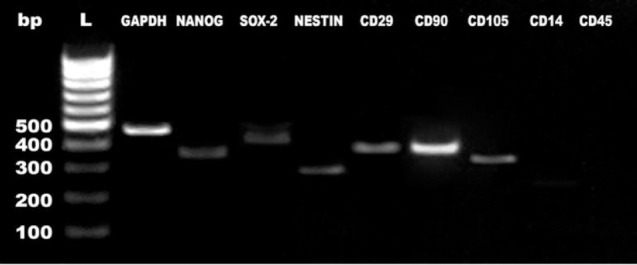
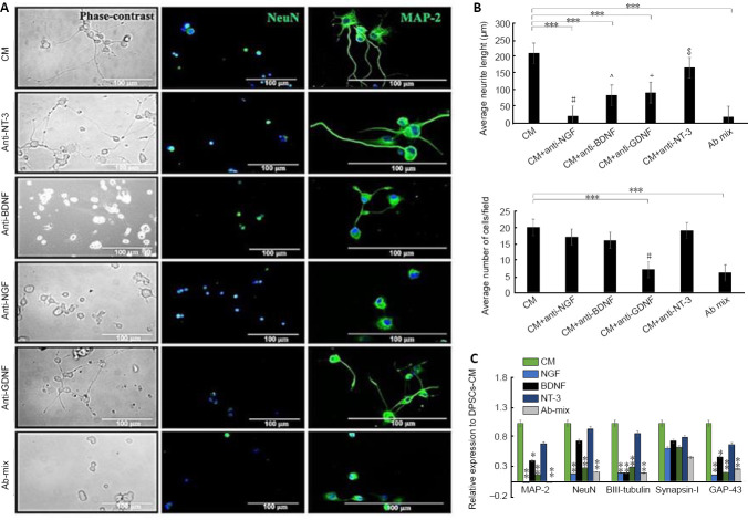
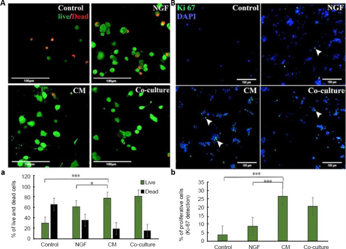
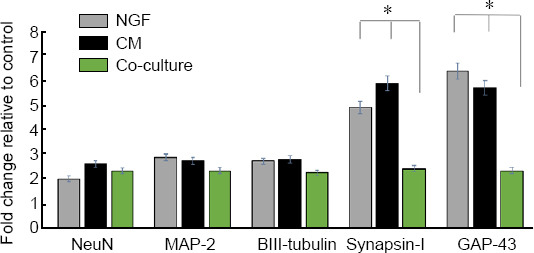
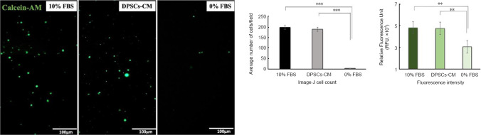
Dental pulp stem cells (DPSCs) secrete neurotrophic factors which may play an important therapeutic role in neural development, maintenance and repair. To test this hypothesis, DPSCs-conditioned medium (DPSCs-CM) was collected from 72 hours serum-free DPSCs cultures. The impact of DPSCs-derived factors on PC12 survival, growth, migration and differentiation was investigated. PC12 cells were treated with nerve growth factor (NGF), DPSCs-CM or co-cultured with DPSCs using Transwell inserts for 8 days. The number of surviving cells with neurite outgrowths and the length of neurites were measured by image analysis. Immunocytochemical staining was used to evaluate the expression of neuronal markers NeuN, microtubule associated protein 2 (MAP-2) and cytoskeletal marker βIII-tubulin. Gene expression levels of axonal growth-associated protein 43 and synaptic protein Synapsin-I, NeuN, MAP-2 and βIII-tubulin were analysed by quantitative polymerase chain reaction (qRT-PCR). DPSCs-CM was analysed for the neurotrophic factors (NGF, brain-derived neurotrophic factor [BDNF], neurotrophin-3, and glial cell-derived neurotrophic factor [GDNF]) by specific ELISAs. Specific neutralizing antibodies against the detected neurotrophic factors were used to study their exact role on PC12 neuronal survival and neurite outgrowth extension. DPSCs-CM significantly promoted cell survival and induced the neurite outgrowth confirmed by NeuN, MAP-2 and βIII-tubulin immunostaining. Furthermore, DPSCs-CM was significantly more effective in stimulating PC12 neurite outgrowths than live DPSCs/PC12 co-cultures over the time studied. The morphology of induced PC12 cells in DPSCs-CM was similar to NGF positive controls; however, DPSCs-CM stimulation of cell survival was significantly higher than what was seen in NGF-treated cultures. The number of surviving PC12 cells treated with DPSCs-CM was markedly reduced by the addition of anti-GDNF, whilst PC12 neurite outgrowth was significantly attenuated by anti-NGF, anti-GDNF and anti-BDNF antibodies. These findings demonstrated that DPSCs were able to promote PC12 survival and differentiation. DPSCs-derived NGF, BDNF and GDNF were involved in the stimulatory action on neurite outgrowth, whereas GDNF also had a significant role in promoting PC12 survival. DPSCs-derived factors may be harnessed as a cell-free therapy for peripheral nerve repair. All experiments were conducted on dead animals that were not sacrificed for the purpose of the study. All the methods were carried out in accordance with Birmingham University guidelines and regulations and the ethical approval is not needed.
牙髓干细胞(DPSCs)分泌神经营养因子,这些因子可能在神经发育、维持和修复中发挥重要的治疗作用。为了验证这一假设,从无血清培养72小时的DPSCs中收集条件培养基(DPSCs-CM)。研究了DPSCs衍生因子对PC12细胞存活、生长、迁移和分化的影响。PC12细胞用神经生长因子(NGF)、DPSCs-CM处理,或使用Transwell小室与DPSCs共培养8天。通过图像分析测量有神经突生长的存活细胞数量和神经突长度。免疫细胞化学染色用于评估神经元标志物NeuN、微管相关蛋白2(MAP-2)和细胞骨架标志物βIII-微管蛋白的表达。通过定量聚合酶链反应(qRT-PCR)分析轴突生长相关蛋白43和突触蛋白Synapsin-I、NeuN、MAP-2和βIII-微管蛋白的基因表达水平。通过特异性酶联免疫吸附测定(ELISA)分析DPSCs-CM中的神经营养因子(NGF、脑源性神经营养因子[BDNF]、神经营养素-3和胶质细胞源性神经营养因子[GDNF])。使用针对检测到的神经营养因子的特异性中和抗体来研究它们对PC12神经元存活和神经突生长延长的确切作用。DPSCs-CM显著促进细胞存活,并通过NeuN、MAP-2和βIII-微管蛋白免疫染色证实诱导神经突生长。此外,在研究期间,DPSCs-CM在刺激PC12神经突生长方面比活的DPSCs/PC12共培养物显著更有效。DPSCs-CM中诱导的PC12细胞形态与NGF阳性对照相似;然而,DPSCs-CM对细胞存活的刺激作用明显高于NGF处理的培养物。添加抗GDNF后,用DPSCs-CM处理的存活PC12细胞数量显著减少,而抗NGF、抗GDNF和抗BDNF抗体显著减弱PC12神经突生长。这些发现表明DPSCs能够促进PC12细胞的存活和分化。DPSCs衍生的NGF、BDNF和GDNF参与了对神经突生长的刺激作用,而GDNF在促进PC12细胞存活方面也具有重要作用。DPSCs衍生因子可作为一种无细胞疗法用于周围神经修复。所有实验均在非为该研究目的而处死的死亡动物身上进行。所有方法均按照伯明翰大学的指导方针和规定进行,无需伦理批准。