Communicable Disease Center (CDC), Hamad Medical Corporation (HMC), Doha, Qatar.
Department of Pediatrics. Hamad Medical Corporation (HMC), Doha, Qatar.
Acta Biomed. 2020 Nov 10;91(4):e2020165. doi: 10.23750/abm.v91i4.10564.
Eosinophils can be considered as multifunctional leukocytes that contribute to various physiological and pathological processes depending on their location and activation status. There are emerging eosinophil-related considerations concerning COVID-19. Variable eosinophil counts have been reported during COVID-19. Whether these changes are related to the primary disease process or due to immunomodulation induced by the treatment has not yet been elucidated.
To describe changes in the differential leukocyte counts including eosinophils, in a cohort of symptomatic patients with confirmed COVID-19 and to correlate these changes, if any, with the severity of the disease.
We recorded the clinical data, lab findings, including inflammatory markers and leukocyte and differential count, course of the disease and severity score in 314 confirmed symptomatic cases of COVID-19.
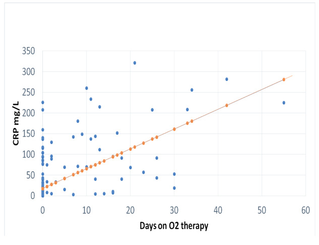
Laboratory tests revealed that 28.7 % (n =86) had mild eosinophilia (eosinophil count > 500 <1,500/µL). Thirty-four patients (11.3%) had elevated absolute neutrophil count (ANC) (>8,000/µL), and 7 (2.3%) had decreased ANC (< 1,500/µl). Seven patients (2.3%) had lymphopenia (<1,000/µL) and 4 (4.67%) had lymphocytosis (> 4,000/µL). C-reactive protein (CRP) was elevated in 83 patients (27.6%). Chest X-Ray changes included: increased broncho vascular markings (38%), ground-glass opacity (GGO) pneumonitis (19.3%), lobar consolidation (5%), bronchopneumonia (8.3%), nodular opacity (1%), acute respiratory distress syndrome (ARDS) (2.3%), pleural effusion (1.0%) and other atypical findings (6.6%). Patients with eosinophilia had significantly lower CRP, and lower % of GGO, lobar and bronchopneumonia and ARDS in their chest images compared to patients without eosinophilia (p: <0.05). They also had a lower requirement for a hospital stay, ICU admission, mechanical ventilation, and oxygen supplementation versus patients without eosinophilia (p: <0.05). The eosinophils count was correlated negatively with the duration of ICU admission, mechanical ventilation, and oxygen supplementation and with CRP level (r: - 0.34, -0.32, -0.61 and - 0.39, respectively) (p: < 0.01).
Our study reports a relatively high prevalence of eosinophilia in symptomatic COVID-19 positive patients. Patients with eosinophilia had a lower level of CRP, milder clinical course and better disease outcomes compared to those without eosinophilia. Our findings indicated a protective role of eosinophils in mitigating the severity of inflammatory diseases through an inhibitory mechanism, as evidenced by lower CRP. This protective role of eosinophils needs to be validated by further prospective studies.
嗜酸性粒细胞可以被认为是多功能白细胞,根据其位置和激活状态,参与各种生理和病理过程。关于 COVID-19,出现了一些与嗜酸性粒细胞相关的新考虑因素。在 COVID-19 期间,报道了嗜酸性粒细胞计数的变化。这些变化是否与主要疾病过程有关,还是由于治疗引起的免疫调节尚不清楚。
描述一组确诊 COVID-19 的有症状患者的白细胞分类计数(包括嗜酸性粒细胞)的变化,并将这些变化与疾病的严重程度相关联。
我们记录了 314 例确诊 COVID-19 有症状患者的临床数据、实验室发现,包括炎症标志物和白细胞及分类计数、疾病过程和严重程度评分。
实验室检查显示,28.7%(n=86)有轻度嗜酸性粒细胞增多症(嗜酸性粒细胞计数>500<1500/µL)。34 例患者(11.3%)有升高的绝对中性粒细胞计数(ANC)(>8000/µL),7 例(2.3%)有降低的 ANC(<1500/µl)。7 例患者(2.3%)有淋巴细胞减少症(<1000/µL),4 例(4.67%)有淋巴细胞增多症(>4000/µL)。83 例患者(27.6%)C 反应蛋白(CRP)升高。胸部 X 线变化包括:支气管血管纹理增加(38%)、磨玻璃影性肺炎(GGO)(19.3%)、肺叶实变(5%)、支气管肺炎(8.3%)、结节状密度影(1%)、急性呼吸窘迫综合征(ARDS)(2.3%)、胸腔积液(1.0%)和其他非典型发现(6.6%)。与无嗜酸性粒细胞增多症的患者相比,有嗜酸性粒细胞增多症的患者 CRP 明显降低,GGO、肺叶和支气管肺炎以及 ARDS 的比例降低(p:<0.05)。他们的住院时间、入住 ICU、机械通气和氧疗需求也低于无嗜酸性粒细胞增多症的患者(p:<0.05)。嗜酸性粒细胞计数与 ICU 住院时间、机械通气和氧疗时间以及 CRP 水平呈负相关(r:-0.34、-0.32、-0.61 和-0.39,分别)(p:<0.01)。
我们的研究报告了 COVID-19 阳性症状患者中嗜酸性粒细胞增多症的相对高发率。与无嗜酸性粒细胞增多症的患者相比,有嗜酸性粒细胞增多症的患者 CRP 水平较低,临床过程较轻,疾病结局较好。我们的研究结果表明,嗜酸性粒细胞通过抑制机制在减轻炎症性疾病的严重程度方面发挥了保护作用,这一点可以通过 CRP 水平降低得到证明。嗜酸性粒细胞的这种保护作用需要通过进一步的前瞻性研究来验证。