Center for Aging and Regeneration, Departamento de Biología Celular y Molecular, Facultad de Ciencias Biológicas, Pontificia Universidad Católica de Chile, Alameda 340, Santiago, Chile.
Laboratorio de Neurobiología Comparada, Instituto Cavanilles, Universidad de Valencia, CIBERNED, 46980, Valencia, Spain.
Neural Dev. 2021 Feb 2;16(1):2. doi: 10.1186/s13064-021-00152-2.
The efficient regenerative abilities at larvae stages followed by a non-regenerative response after metamorphosis in froglets makes Xenopus an ideal model organism to understand the cellular responses leading to spinal cord regeneration.
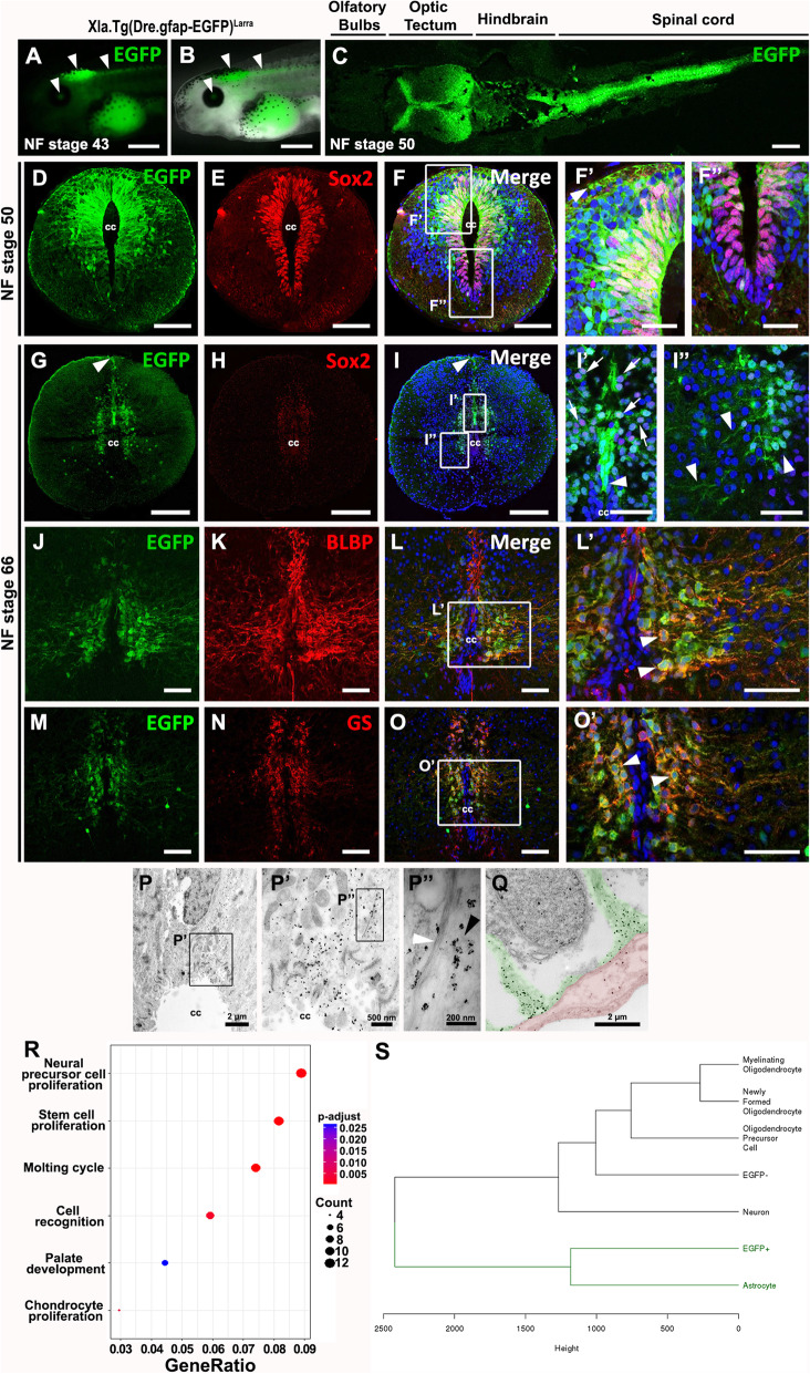
We compared the cellular response to spinal cord injury between the regenerative and non-regenerative stages of Xenopus laevis. For this analysis, we used electron microscopy, immunofluorescence and histological staining of the extracellular matrix. We generated two transgenic lines: i) the reporter line with the zebrafish GFAP regulatory regions driving the expression of EGFP, and ii) a cell specific inducible ablation line with the same GFAP regulatory regions. In addition, we used FACS to isolate EGFP cells for RNAseq analysis.
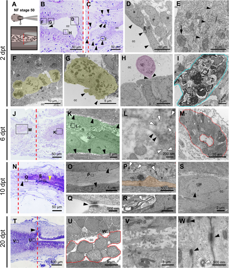
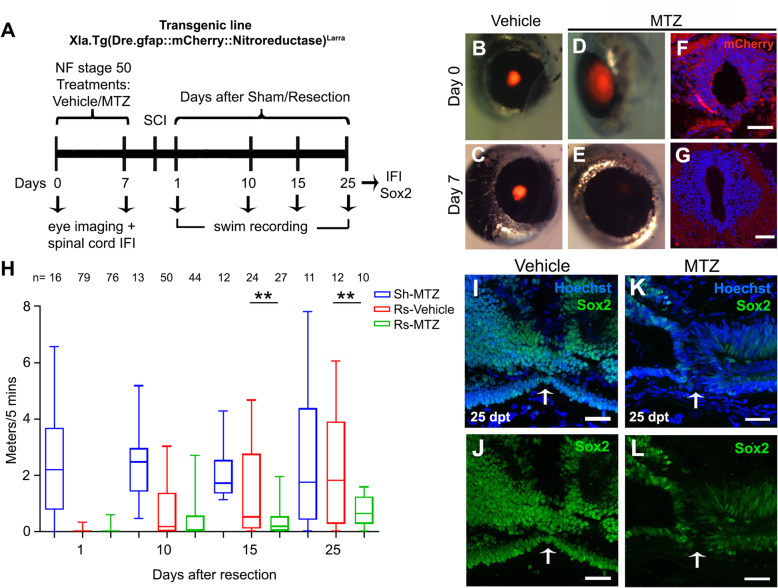
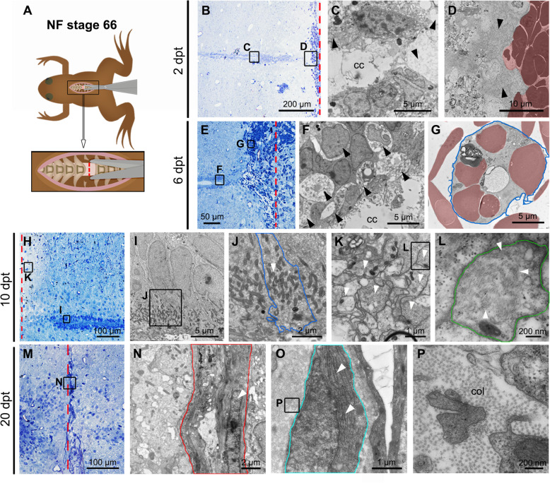
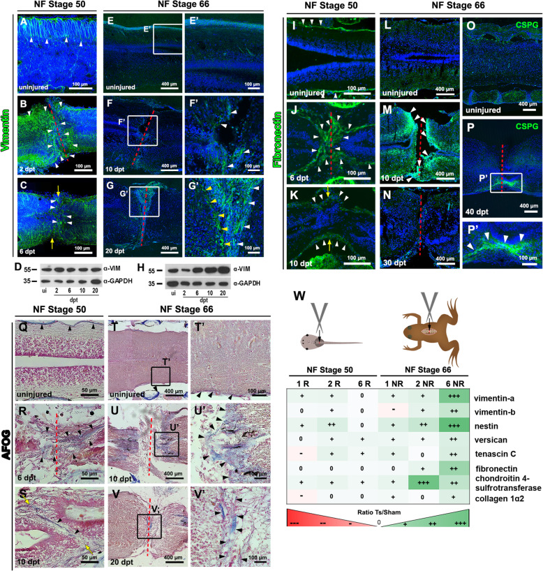
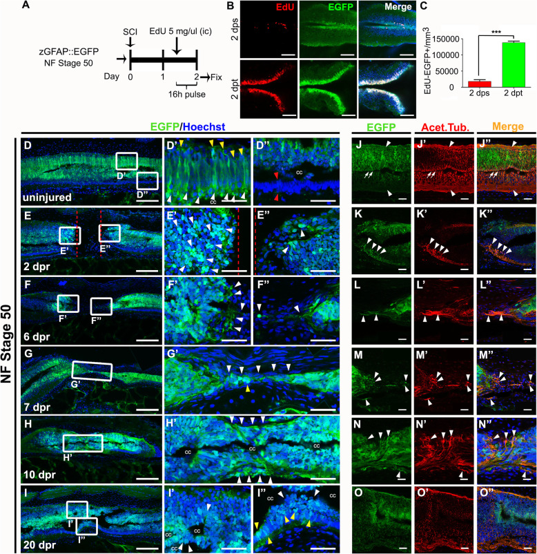
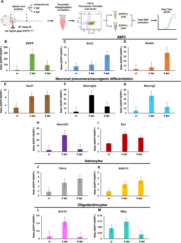
In regenerative stage animals, spinal cord regeneration triggers a rapid sealing of the injured stumps, followed by proliferation of cells lining the central canal, and formation of rosette-like structures in the ablation gap. In addition, the central canal is filled by cells with similar morphology to the cells lining the central canal, neurons, axons, and even synaptic structures. Regeneration is almost completed after 20 days post injury. In non-regenerative stage animals, mostly damaged tissue was observed, without clear closure of the stumps. The ablation gap was filled with fibroblast-like cells, and deposition of extracellular matrix components. No reconstruction of the spinal cord was observed even after 40 days post injury. Cellular markers analysis confirmed these histological differences, a transient increase of vimentin, fibronectin and collagen was detected in regenerative stages, contrary to a sustained accumulation of most of these markers, including chondroitin sulfate proteoglycans in the NR-stage. The zebrafish GFAP transgenic line was validated, and we have demonstrated that is a very reliable and new tool to study the role of neural stem progenitor cells (NSPCs). RNASeq of GFAP::EGFP cells has allowed us to clearly demonstrate that indeed these cells are NSPCs. On the contrary, the GFAP::EGFP transgene is mainly expressed in astrocytes in non-regenerative stages. During regenerative stages, spinal cord injury activates proliferation of NSPCs, and we found that are mainly differentiated into neurons and glial cells. Specific ablation of these cells abolished proper regeneration, confirming that NSPCs cells are necessary for functional regeneration of the spinal cord.
The cellular response to spinal cord injury in regenerative and non-regenerative stages is profoundly different between both stages. A key hallmark of the regenerative response is the activation of NSPCs, which massively proliferate, and are differentiated into neurons to reconstruct the spinal cord. Also very notably, no glial scar formation is observed in regenerative stages, but a transient, glial scar-like structure is formed in non-regenerative stage animals.
在蛙类幼虫阶段具有高效的再生能力,而在变态后则表现出非再生反应,这使得非洲爪蟾成为研究导致脊髓再生的细胞反应的理想模式生物。
我们比较了再生和非再生阶段非洲爪蟾的脊髓损伤的细胞反应。为此分析,我们使用了电子显微镜、免疫荧光和细胞外基质的组织学染色。我们生成了两个转基因系:i)具有斑马鱼 GFAP 调节区驱动 EGFP 表达的报告系,和 ii)具有相同 GFAP 调节区的细胞特异性诱导消融系。此外,我们使用 FACS 分离 EGFP 细胞进行 RNAseq 分析。
在再生阶段动物中,脊髓再生引发受伤残端的快速封闭,随后是中央管衬里细胞的增殖,并在消融间隙中形成玫瑰花结样结构。此外,中央管被与中央管衬里细胞、神经元、轴突甚至突触结构相似形态的细胞填充。损伤后 20 天几乎完成了再生。在非再生阶段动物中,主要观察到受损组织,残端没有明显的封闭。消融间隙充满了成纤维细胞样细胞和细胞外基质成分的沉积。即使在损伤后 40 天,也没有观察到脊髓的重建。细胞标志物分析证实了这些组织学差异,在再生阶段检测到波形蛋白、纤维连接蛋白和胶原蛋白的短暂增加,而在非再生阶段则检测到大多数这些标志物(包括软骨素硫酸蛋白聚糖)的持续积累。验证了斑马鱼 GFAP 转基因系,并且我们已经证明它是研究神经干细胞祖细胞 (NSPCs) 作用的非常可靠和新工具。GFAP::EGFP 细胞的 RNAseq 允许我们清楚地证明这些细胞确实是 NSPCs。相反,GFAP::EGFP 转基因主要在非再生阶段的星形胶质细胞中表达。在再生阶段,脊髓损伤激活 NSPCs 的增殖,我们发现它们主要分化为神经元和神经胶质细胞。这些细胞的特异性消融消除了适当的再生,证实 NSPCs 细胞对于脊髓的功能再生是必需的。
再生和非再生阶段脊髓损伤的细胞反应在两个阶段之间有很大的不同。再生反应的一个关键标志是 NSPCs 的激活,它大量增殖,并分化为神经元以重建脊髓。同样值得注意的是,在再生阶段没有观察到神经胶质瘢痕形成,而是在非再生阶段动物中形成短暂的、神经胶质瘢痕样结构。