• 文献检索
  • 文档翻译
  • 深度研究
  • 学术资讯
  • Suppr Zotero 插件Zotero 插件
  • 邀请有礼
  • 套餐&价格
  • 历史记录
应用&插件
Suppr Zotero 插件Zotero 插件浏览器插件Mac 客户端Windows 客户端微信小程序
定价
高级版会员购买积分包购买API积分包
服务
文献检索文档翻译深度研究API 文档MCP 服务
关于我们
关于 Suppr公司介绍联系我们用户协议隐私条款
关注我们

Suppr 超能文献

核心技术专利:CN118964589B侵权必究
粤ICP备2023148730 号-1Suppr @ 2026

文献检索

告别复杂PubMed语法,用中文像聊天一样搜索,搜遍4000万医学文献。AI智能推荐,让科研检索更轻松。

立即免费搜索

文件翻译

保留排版,准确专业,支持PDF/Word/PPT等文件格式,支持 12+语言互译。

免费翻译文档

深度研究

AI帮你快速写综述,25分钟生成高质量综述,智能提取关键信息,辅助科研写作。

立即免费体验

结合体内角膜共焦显微镜与基于深度学习的分析揭示急性猴免疫缺陷病毒感染中的感觉神经纤维损失。

Combining In Vivo Corneal Confocal Microscopy With Deep Learning-Based Analysis Reveals Sensory Nerve Fiber Loss in Acute Simian Immunodeficiency Virus Infection.

机构信息

Department of Molecular and Comparative Pathobiology, Johns Hopkins University School of Medicine, Baltimore, MD.

Departments of Epidemiology and Biostatistics, Johns Hopkins Bloomberg School of Public Health, Baltimore, MD.

出版信息

Cornea. 2021 May 1;40(5):635-642. doi: 10.1097/ICO.0000000000002661.

DOI:10.1097/ICO.0000000000002661
PMID:33528225
原文链接:https://pmc.ncbi.nlm.nih.gov/articles/PMC8009813/
Abstract

PURPOSE

To characterize corneal subbasal nerve plexus features of normal and simian immunodeficiency virus (SIV)-infected macaques by combining in vivo corneal confocal microscopy (IVCM) with automated assessments using deep learning-based methods customized for macaques.

METHODS

IVCM images were collected from both male and female age-matched rhesus and pigtailed macaques housed at the Johns Hopkins University breeding colony using the Heidelberg HRTIII with Rostock Corneal Module. We also obtained repeat IVCM images of 12 SIV-infected animals including preinfection and 10-day post-SIV infection time points. All IVCM images were analyzed using a deep convolutional neural network architecture developed specifically for macaque studies.

RESULTS

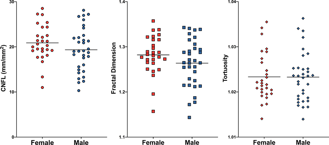
Deep learning-based segmentation of subbasal nerves in IVCM images from macaques demonstrated that corneal nerve fiber length and fractal dimension measurements did not differ between species, but pigtailed macaques had significantly higher baseline corneal nerve fiber tortuosity than rhesus macaques (P = 0.005). Neither sex nor age of macaques was associated with differences in any of the assessed corneal subbasal nerve parameters. In the SIV/macaque model of human immunodeficiency virus, acute SIV infection induced significant decreases in both corneal nerve fiber length and fractal dimension (P = 0.01 and P = 0.008, respectively).

CONCLUSIONS

The combination of IVCM and robust objective deep learning analysis is a powerful tool to track sensory nerve damage, enabling early detection of neuropathy. Adapting deep learning analyses to clinical corneal nerve assessments will improve monitoring of small sensory nerve fiber damage in numerous clinical settings including human immunodeficiency virus.

摘要

目的

通过将活体角膜共聚焦显微镜(IVCM)与基于深度学习的专为猕猴定制的自动评估方法相结合,来描述正常和猴免疫缺陷病毒(SIV)感染猕猴的角膜基底下神经丛特征。

方法

使用海德堡 HRTIII 与罗斯托克角膜模块,从约翰霍普金斯大学繁殖群中年龄匹配的雄性和雌性恒河猴和长尾猕猴中采集 IVCM 图像。我们还获得了 12 只 SIV 感染动物的重复 IVCM 图像,包括感染前和感染后 10 天的时间点。所有 IVCM 图像均使用专为猕猴研究开发的深度卷积神经网络架构进行分析。

结果

基于深度学习的猕猴 IVCM 图像中基底下神经的分割表明,种间角膜神经纤维长度和分形维数测量值没有差异,但长尾猕猴的角膜神经纤维扭曲度明显高于恒河猴(P=0.005)。猕猴的性别或年龄均与评估的任何角膜基底下神经参数无差异相关。在 SIV/猕猴人类免疫缺陷病毒模型中,急性 SIV 感染导致角膜神经纤维长度和分形维数均显著下降(分别为 P=0.01 和 P=0.008)。

结论

IVCM 与强大的客观深度学习分析相结合是一种跟踪感觉神经损伤的有力工具,能够早期发现神经病变。将深度学习分析应用于临床角膜神经评估将改善对包括人类免疫缺陷病毒在内的众多临床环境中小感觉神经纤维损伤的监测。

https://cdn.ncbi.nlm.nih.gov/pmc/blobs/5e66/8009813/2b480d8bdded/nihms-1653938-f0007.jpg
https://cdn.ncbi.nlm.nih.gov/pmc/blobs/5e66/8009813/39b519938a98/nihms-1653938-f0001.jpg
https://cdn.ncbi.nlm.nih.gov/pmc/blobs/5e66/8009813/cf8d6e015f12/nihms-1653938-f0002.jpg
https://cdn.ncbi.nlm.nih.gov/pmc/blobs/5e66/8009813/0bb781d0e4f0/nihms-1653938-f0003.jpg
https://cdn.ncbi.nlm.nih.gov/pmc/blobs/5e66/8009813/2feecbd0a669/nihms-1653938-f0004.jpg
https://cdn.ncbi.nlm.nih.gov/pmc/blobs/5e66/8009813/41f220d88a93/nihms-1653938-f0005.jpg
https://cdn.ncbi.nlm.nih.gov/pmc/blobs/5e66/8009813/4d32f3dbcd5a/nihms-1653938-f0006.jpg
https://cdn.ncbi.nlm.nih.gov/pmc/blobs/5e66/8009813/2b480d8bdded/nihms-1653938-f0007.jpg

相似文献

1
Combining In Vivo Corneal Confocal Microscopy With Deep Learning-Based Analysis Reveals Sensory Nerve Fiber Loss in Acute Simian Immunodeficiency Virus Infection.结合体内角膜共焦显微镜与基于深度学习的分析揭示急性猴免疫缺陷病毒感染中的感觉神经纤维损失。
Cornea. 2021 May 1;40(5):635-642. doi: 10.1097/ICO.0000000000002661.
2
Loss of corneal sensory nerve fibers in SIV-infected macaques: an alternate approach to investigate HIV-induced PNS damage.感染猴免疫缺陷病毒(SIV)的猕猴角膜感觉神经纤维的丧失:一种研究人类免疫缺陷病毒(HIV)诱导的周围神经系统损伤的替代方法。
Am J Pathol. 2014 Jun;184(6):1652-9. doi: 10.1016/j.ajpath.2014.02.009. Epub 2014 May 12.
3
Persistent Peripheral Nervous System Damage in Simian Immunodeficiency Virus-Infected Macaques Receiving Antiretroviral Therapy.接受抗逆转录病毒治疗的感染猿猴免疫缺陷病毒的猕猴存在持续性外周神经系统损伤
J Neuropathol Exp Neurol. 2015 Nov;74(11):1053-60. doi: 10.1097/NEN.0000000000000249.
4
A Deep Learning Model for Automated Sub-Basal Corneal Nerve Segmentation and Evaluation Using In Vivo Confocal Microscopy.一种用于使用活体共聚焦显微镜进行自动化角膜基底膜下神经分割和评估的深度学习模型。
Transl Vis Sci Technol. 2020 Jun 18;9(2):32. doi: 10.1167/tvst.9.2.32. eCollection 2020 Jun.
5
In vivo confocal microscopy morphometric analysis of corneal subbasal nerve plexus in dry eye disease using newly developed fully automated system.使用新开发的全自动系统对干眼症患者角膜基底神经丛进行体内共焦显微镜形态计量分析。
Graefes Arch Clin Exp Ophthalmol. 2019 Mar;257(3):583-589. doi: 10.1007/s00417-018-04225-7. Epub 2019 Jan 14.
6
Degeneration and Regeneration of Subbasal Corneal Nerves after Infectious Keratitis: A Longitudinal In Vivo Confocal Microscopy Study.感染性角膜炎后角膜基底膜下神经的退变与再生:一项纵向活体共聚焦显微镜研究
Ophthalmology. 2015 Nov;122(11):2200-9. doi: 10.1016/j.ophtha.2015.06.047. Epub 2015 Aug 6.
7
Generative Adversarial Network Based Automatic Segmentation of Corneal Subbasal Nerves on In Vivo Confocal Microscopy Images.基于生成对抗网络的活体共聚焦显微镜图像下角膜基底部神经的自动分割。
Transl Vis Sci Technol. 2021 May 3;10(6):33. doi: 10.1167/tvst.10.6.33.
8
Linking pig-tailed macaque major histocompatibility complex class I haplotypes and cytotoxic T lymphocyte escape mutations in simian immunodeficiency virus infection.将猪尾猕猴主要组织相容性复合体I类单倍型与猿猴免疫缺陷病毒感染中的细胞毒性T淋巴细胞逃逸突变联系起来。
J Virol. 2014 Dec;88(24):14310-25. doi: 10.1128/JVI.02428-14. Epub 2014 Oct 1.
9
Macaque species susceptibility to simian immunodeficiency virus: increased incidence of SIV central nervous system disease in pigtailed macaques versus rhesus macaques.猕猴物种对猿猴免疫缺陷病毒的易感性:与恒河猴相比,猪尾猕猴感染猿猴免疫缺陷病毒后中枢神经系统疾病的发病率增加。
J Neurovirol. 2015 Apr;21(2):148-58. doi: 10.1007/s13365-015-0313-7. Epub 2015 Feb 12.
10
In vivo confocal microscopy study of corneal nerve alterations in children and youths with Type 1 diabetes.1 型糖尿病患儿和青少年角膜神经改变的活体共聚焦显微镜研究。
Pediatr Diabetes. 2021 Aug;22(5):780-786. doi: 10.1111/pedi.13219. Epub 2021 May 11.

引用本文的文献

1
Multimodal Approach in Dry Eye Disease Combining In Vivo Confocal Microscopy and HLA-DR Expression.多模态方法在干眼症中的应用,结合了活体共聚焦显微镜和 HLA-DR 表达。
Transl Vis Sci Technol. 2024 Aug 1;13(8):39. doi: 10.1167/tvst.13.8.39.
2
A Switch from Glial to Neuronal Gene Expression Alterations in the Spinal Cord of SIV-infected Macaques on Antiretroviral Therapy.在接受抗逆转录病毒治疗的 SIV 感染猕猴的脊髓中,从神经胶质到神经元的基因表达变化的转变。
J Neuroimmune Pharmacol. 2024 Jun 12;19(1):28. doi: 10.1007/s11481-024-10130-0.
3
Potential applications of artificial intelligence in image analysis in cornea diseases: a review.
人工智能在角膜疾病图像分析中的潜在应用:综述
Eye Vis (Lond). 2024 Mar 7;11(1):10. doi: 10.1186/s40662-024-00376-3.
4
Evaluation of a computer-aided diagnostic model for corneal diseases by analyzing confocal microscopy images.通过分析共焦显微镜图像评估角膜疾病的计算机辅助诊断模型
Front Med (Lausanne). 2023 Apr 20;10:1164188. doi: 10.3389/fmed.2023.1164188. eCollection 2023.
5
Corneal Confocal Microscopy as a Quantitative Imaging Biomarker of Diabetic Peripheral Neuropathy: A Review.角膜共聚焦显微镜作为糖尿病周围神经病变的定量成像生物标志物:综述
J Clin Med. 2022 Aug 31;11(17):5130. doi: 10.3390/jcm11175130.
6
Corneal Nerve Changes Observed by In Vivo Confocal Microscopy in Patients Receiving Oxaliplatin for Colorectal Cancer: The COCO Study.通过活体共聚焦显微镜观察接受奥沙利铂治疗的结直肠癌患者的角膜神经变化:COCO研究
J Clin Med. 2022 Aug 16;11(16):4770. doi: 10.3390/jcm11164770.