Koç University Research Center for Translational Medicine, Koç University, Istanbul, Turkey.
Techy Bilişim Ltd., Eskişehir, Turkey.
Transl Vis Sci Technol. 2021 May 3;10(6):33. doi: 10.1167/tvst.10.6.33.
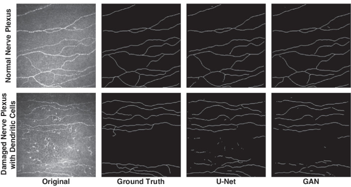
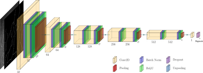
In vivo confocal microscopy (IVCM) is a noninvasive, reproducible, and inexpensive diagnostic tool for corneal diseases. However, widespread and effortless image acquisition in IVCM creates serious image analysis workloads on ophthalmologists, and neural networks could solve this problem quickly. We have produced a novel deep learning algorithm based on generative adversarial networks (GANs), and we compare its accuracy for automatic segmentation of subbasal nerves in IVCM images with a fully convolutional neural network (U-Net) based method.
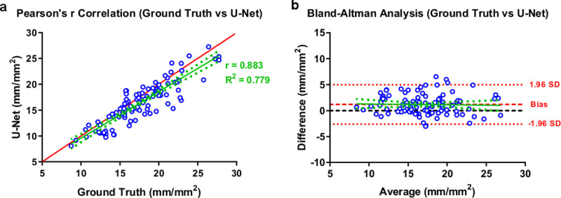
We have collected IVCM images from 85 subjects. U-Net and GAN-based image segmentation methods were trained and tested under the supervision of three clinicians for the segmentation of corneal subbasal nerves. Nerve segmentation results for GAN and U-Net-based methods were compared with the clinicians by using Pearson's R correlation, Bland-Altman analysis, and receiver operating characteristics (ROC) statistics. Additionally, different noises were applied on IVCM images to evaluate the performances of the algorithms with noises of biomedical imaging.
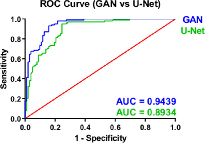
The GAN-based algorithm demonstrated similar correlation and Bland-Altman analysis results with U-Net. The GAN-based method showed significantly higher accuracy compared to U-Net in ROC curves. Additionally, the performance of the U-Net deteriorated significantly with different noises, especially in speckle noise, compared to GAN.
This study is the first application of GAN-based algorithms on IVCM images. The GAN-based algorithms demonstrated higher accuracy than U-Net for automatic corneal nerve segmentation in IVCM images, in patient-acquired images and noise applied images. This GAN-based segmentation method can be used as a facilitating diagnostic tool in ophthalmology clinics.
Generative adversarial networks are emerging deep learning models for medical image processing, which could be important clinical tools for rapid segmentation and analysis of corneal subbasal nerves in IVCM images.
共焦激光角膜显微镜(IVCM)是一种非侵入性、可重复性和廉价的角膜疾病诊断工具。然而,IVCM 中广泛且轻松的图像采集给眼科医生带来了严重的图像分析工作量,而神经网络可以快速解决这个问题。我们开发了一种基于生成对抗网络(GAN)的新型深度学习算法,并比较了该算法在自动分割 IVCM 图像中基底下神经方面的准确性与基于全卷积神经网络(U-Net)的方法。
我们从 85 名患者中收集了 IVCM 图像。U-Net 和基于 GAN 的图像分割方法在三位临床医生的监督下进行了训练和测试,以分割角膜基底下神经。通过 Pearson R 相关性、Bland-Altman 分析和受试者工作特征(ROC)统计,将 GAN 和基于 U-Net 的方法的神经分割结果与临床医生进行了比较。此外,还在 IVCM 图像上应用了不同的噪声,以评估算法在生物医学成像噪声下的性能。
基于 GAN 的算法与 U-Net 具有相似的相关性和 Bland-Altman 分析结果。与 U-Net 相比,基于 GAN 的方法在 ROC 曲线中表现出更高的准确性。此外,与 GAN 相比,U-Net 在不同噪声下的性能明显恶化,尤其是在斑点噪声下。
这是首次将基于 GAN 的算法应用于 IVCM 图像。基于 GAN 的算法在自动角膜神经分割方面的准确性优于 U-Net,无论是在患者采集的图像还是在应用噪声的图像中。这种基于 GAN 的分割方法可以作为眼科临床的一种辅助诊断工具。
王春旭