• 文献检索
  • 文档翻译
  • 深度研究
  • 学术资讯
  • Suppr Zotero 插件Zotero 插件
  • 邀请有礼
  • 套餐&价格
  • 历史记录
应用&插件
Suppr Zotero 插件Zotero 插件浏览器插件Mac 客户端Windows 客户端微信小程序
定价
高级版会员购买积分包购买API积分包
服务
文献检索文档翻译深度研究API 文档MCP 服务
关于我们
关于 Suppr公司介绍联系我们用户协议隐私条款
关注我们

Suppr 超能文献

核心技术专利:CN118964589B侵权必究
粤ICP备2023148730 号-1Suppr @ 2026

文献检索

告别复杂PubMed语法,用中文像聊天一样搜索,搜遍4000万医学文献。AI智能推荐,让科研检索更轻松。

立即免费搜索

文件翻译

保留排版,准确专业,支持PDF/Word/PPT等文件格式,支持 12+语言互译。

免费翻译文档

深度研究

AI帮你快速写综述,25分钟生成高质量综述,智能提取关键信息,辅助科研写作。

立即免费体验

中风神经影像学中的放射组学:技术、应用与挑战。

Radiomics in Stroke Neuroimaging: Techniques, Applications, and Challenges.

作者信息

Chen Qian, Xia Tianyi, Zhang Mingyue, Xia Nengzhi, Liu Jinjin, Yang Yunjun

机构信息

Department of Radiology, The First Affiliated Hospital of Wenzhou Medical University, Zhejiang, China.

出版信息

Aging Dis. 2021 Feb 1;12(1):143-154. doi: 10.14336/AD.2020.0421. eCollection 2021 Feb.

DOI:10.14336/AD.2020.0421
PMID:33532134
原文链接:https://pmc.ncbi.nlm.nih.gov/articles/PMC7801280/
Abstract

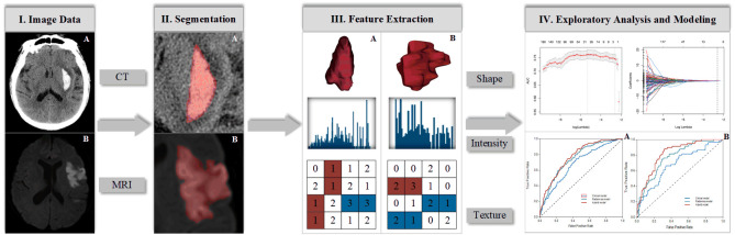
Stroke is a leading cause of disability and mortality worldwide, resulting in substantial economic costs for post-stroke care each year. Neuroimaging, such as cranial computed tomography or magnetic resonance imaging, is the backbone of stroke management strategies, which can guide treatment decision-making (thrombolysis or hemostasis) at an early stage. With advances in computational technologies, particularly in machine learning, visual image information can now be converted into numerous quantitative features in an objective, repeatable, and high-throughput manner, in a process known as radiomics. Radiomics is mainly used in the field of oncology, which remains an area of active research. Over the past few years, investigators have attempted to apply radiomics to stroke in the hope of gaining benefits similar to those obtained in cancer management, i.e., in promoting the development of personalized precision medicine. Currently, radiomic analysis has shown promise for a variety of applications in stroke, including the diagnosis of stroke lesions, early prediction of outcomes, and evaluation for long-term prognosis. In this article, we elaborate the contributions of radiomics to stroke, as well as the subprocesses and techniques involved in radiomics studies. We also discuss the potential challenges facing its widespread implementation in routine practice and the directions for future research.

摘要

中风是全球残疾和死亡的主要原因,每年给中风后护理带来巨大的经济成本。神经影像学,如头颅计算机断层扫描或磁共振成像,是中风管理策略的支柱,可在早期阶段指导治疗决策(溶栓或止血)。随着计算技术的进步,特别是机器学习领域的进步,视觉图像信息现在可以以客观、可重复和高通量的方式转换为大量定量特征,这一过程称为放射组学。放射组学主要应用于肿瘤学领域,该领域仍是一个活跃的研究领域。在过去几年中,研究人员试图将放射组学应用于中风,希望获得与癌症管理类似的益处,即促进个性化精准医学的发展。目前,放射组学分析在中风的各种应用中显示出前景,包括中风病变的诊断、结局的早期预测和长期预后评估。在本文中,我们阐述了放射组学对中风的贡献,以及放射组学研究中涉及的子过程和技术。我们还讨论了其在常规实践中广泛应用面临的潜在挑战以及未来研究的方向。

https://cdn.ncbi.nlm.nih.gov/pmc/blobs/bc1b/7801280/d9626fa9644e/ad-12-1-143-g2.jpg
https://cdn.ncbi.nlm.nih.gov/pmc/blobs/bc1b/7801280/e6c891bfd49e/ad-12-1-143-g1.jpg
https://cdn.ncbi.nlm.nih.gov/pmc/blobs/bc1b/7801280/d9626fa9644e/ad-12-1-143-g2.jpg
https://cdn.ncbi.nlm.nih.gov/pmc/blobs/bc1b/7801280/e6c891bfd49e/ad-12-1-143-g1.jpg
https://cdn.ncbi.nlm.nih.gov/pmc/blobs/bc1b/7801280/d9626fa9644e/ad-12-1-143-g2.jpg

相似文献

1
Radiomics in Stroke Neuroimaging: Techniques, Applications, and Challenges.中风神经影像学中的放射组学:技术、应用与挑战。
Aging Dis. 2021 Feb 1;12(1):143-154. doi: 10.14336/AD.2020.0421. eCollection 2021 Feb.
2
MRI Radiomics and Predictive Models in Assessing Ischemic Stroke Outcome-A Systematic Review.MRI影像组学与预测模型在评估缺血性脑卒中预后中的应用——一项系统综述
Diagnostics (Basel). 2023 Feb 23;13(5):857. doi: 10.3390/diagnostics13050857.
3
Radiomics in stratification of pancreatic cystic lesions: Machine learning in action.基于影像组学的胰腺囊性病变危险分层:机器学习的实践。
Cancer Lett. 2020 Jan 28;469:228-237. doi: 10.1016/j.canlet.2019.10.023. Epub 2019 Oct 17.
4
Potential and limitations of radiomics in neuro-oncology.放射组学在神经肿瘤学中的潜力和局限性。
J Clin Neurosci. 2021 Aug;90:206-211. doi: 10.1016/j.jocn.2021.05.015. Epub 2021 Jun 11.
5
Radiomics: the bridge between medical imaging and personalized medicine.放射组学:医学影像与个性化医疗之间的桥梁。
Nat Rev Clin Oncol. 2017 Dec;14(12):749-762. doi: 10.1038/nrclinonc.2017.141. Epub 2017 Oct 4.
6
Radiomics in Breast Imaging from Techniques to Clinical Applications: A Review.乳腺影像中的放射组学:从技术到临床应用综述。
Korean J Radiol. 2020 Jul;21(7):779-792. doi: 10.3348/kjr.2019.0855.
7
A review in radiomics: Making personalized medicine a reality via routine imaging.放射组学综述:通过常规成像实现个体化医疗。
Med Res Rev. 2022 Jan;42(1):426-440. doi: 10.1002/med.21846. Epub 2021 Jul 26.
8
Radiomics approaches in gastric cancer: a frontier in clinical decision making.胃癌的放射组学方法:临床决策制定的前沿领域。
Chin Med J (Engl). 2019 Aug 20;132(16):1983-1989. doi: 10.1097/CM9.0000000000000360.
9
Novel imaging techniques of rectal cancer: what do radiomics and radiogenomics have to offer? A literature review.直肠癌的新型影像学技术:放射组学和放射基因组学有何贡献?文献综述。
Abdom Radiol (NY). 2019 Nov;44(11):3764-3774. doi: 10.1007/s00261-019-02042-y.
10
Cardiac computed tomography radiomics: a narrative review of current status and future directions.心脏计算机断层扫描放射组学:现状与未来方向的叙述性综述
Quant Imaging Med Surg. 2022 Jun;12(6):3436-3453. doi: 10.21037/qims-21-1022.

引用本文的文献

1
Predicting malignant cerebral edema after acute ischemic stroke: a machine-learning model with multi-region radiomics.预测急性缺血性卒中后恶性脑水肿:一种基于多区域放射组学的机器学习模型
Quant Imaging Med Surg. 2025 Jun 6;15(6):5188-5203. doi: 10.21037/qims-2024-2751. Epub 2025 Jun 3.
2
Accuracy of Machine Learning in Predicting Post-Stroke Depression: A Systematic Review and Meta-Analysis.机器学习预测中风后抑郁的准确性:一项系统评价与荟萃分析。
Brain Behav. 2025 May;15(5):e70557. doi: 10.1002/brb3.70557.
3
Diffusion-Weighted Imaging-Based Radiomics Features and Machine Learning Method to Predict the 90-Day Prognosis in Patients With Acute Ischemic Stroke.

本文引用的文献

1
Penumbra-based radiomics signature as prognostic biomarkers for thrombolysis of acute ischemic stroke patients: a multicenter cohort study.基于半影区的放射组学特征作为急性缺血性脑卒中溶栓患者预后生物标志物的研究:一项多中心队列研究。
J Neurol. 2020 May;267(5):1454-1463. doi: 10.1007/s00415-020-09713-7. Epub 2020 Feb 1.
2
Texture Features of Magnetic Resonance Images: an Early Marker of Post-stroke Cognitive Impairment.磁共振图像纹理特征:中风后认知障碍的早期标志物。
Transl Stroke Res. 2020 Aug;11(4):643-652. doi: 10.1007/s12975-019-00746-3. Epub 2019 Nov 1.
3
Reproducibility of CT Radiomic Features within the Same Patient: Influence of Radiation Dose and CT Reconstruction Settings.
基于扩散加权成像的放射组学特征及机器学习方法预测急性缺血性脑卒中患者90天预后
Neurologist. 2025 Mar 1;30(2):93-101. doi: 10.1097/NRL.0000000000000599.
4
Functional Connectivity and MRI Radiomics Biomarkers of Cognitive and Brain Reserve in Post-Stroke Cognitive Impairment Prediction-A Study Protocol.用于中风后认知障碍预测的认知与脑储备的功能连接性和MRI影像组学生物标志物——一项研究方案
Life (Basel). 2025 Jan 20;15(1):131. doi: 10.3390/life15010131.
5
Clinical-Radiomics Nomogram Model Based on CT Angiography for Prediction of Intracranial Aneurysm Rupture: A Multicenter Study.基于CT血管造影的临床-影像组学列线图模型预测颅内动脉瘤破裂:一项多中心研究
J Multidiscip Healthc. 2024 Dec 10;17:5917-5926. doi: 10.2147/JMDH.S491697. eCollection 2024.
6
Research on imaging biomarkers for chronic subdural hematoma recurrence.慢性硬膜下血肿复发的影像学生物标志物研究。
Med Biol Eng Comput. 2025 Mar;63(3):823-834. doi: 10.1007/s11517-024-03232-7. Epub 2024 Nov 6.
7
Machine learning-based radiomics in neurodegenerative and cerebrovascular disease.基于机器学习的神经退行性疾病和脑血管疾病的影像组学
MedComm (2020). 2024 Oct 28;5(11):e778. doi: 10.1002/mco2.778. eCollection 2024 Nov.
8
Radiomics in radiology: What the radiologist needs to know about technical aspects and clinical impact.放射学中的放射组学:放射科医生需要了解的技术方面和临床影响。
Radiol Med. 2024 Dec;129(12):1751-1765. doi: 10.1007/s11547-024-01904-w. Epub 2024 Oct 30.
9
Predictive value of radiomics for intracranial aneurysm rupture: a systematic review and meta-analysis.放射组学对颅内动脉瘤破裂的预测价值:一项系统评价和荟萃分析。
Front Neurosci. 2024 Oct 9;18:1474780. doi: 10.3389/fnins.2024.1474780. eCollection 2024.
10
A machine learning model based on results of a comprehensive radiological evaluation can predict the prognosis of basal ganglia cerebral hemorrhage treated with neuroendoscopy.基于全面放射学评估结果的机器学习模型可以预测神经内镜治疗基底节区脑出血的预后。
Front Neurol. 2024 Oct 1;15:1406271. doi: 10.3389/fneur.2024.1406271. eCollection 2024.
同一患者内 CT 放射组特征的可重复性:辐射剂量和 CT 重建参数的影响。
Radiology. 2019 Dec;293(3):583-591. doi: 10.1148/radiol.2019190928. Epub 2019 Oct 1.
4
Early prediction of clinical outcomes in patients with aneurysmal subarachnoid hemorrhage using computed tomography texture analysis.利用 CT 纹理分析对颅内动脉瘤性蛛网膜下腔出血患者的临床结局进行早期预测。
J Clin Neurosci. 2020 Jan;71:144-149. doi: 10.1016/j.jocn.2019.08.098. Epub 2019 Sep 4.
5
Radiomics features on non-contrast computed tomography predict early enlargement of spontaneous intracerebral hemorrhage.非增强计算机断层扫描上的影像组学特征可预测自发性脑出血的早期扩大。
Clin Neurol Neurosurg. 2019 Oct;185:105491. doi: 10.1016/j.clineuro.2019.105491. Epub 2019 Aug 15.
6
Noncontrast computer tomography-based radiomics model for predicting intracerebral hemorrhage expansion: preliminary findings and comparison with conventional radiological model.基于非对比计算机断层扫描的放射组学模型预测脑出血扩大:初步研究结果及与传统影像学模型的比较。
Eur Radiol. 2020 Jan;30(1):87-98. doi: 10.1007/s00330-019-06378-3. Epub 2019 Aug 5.
7
Computerized Tomography Radiomics Features Analysis for Evaluation of Perihematomal Edema in Basal Ganglia Hemorrhage.计算机断层扫描影像组学特征分析用于评估基底节区脑出血周围血肿水肿
J Craniofac Surg. 2019 Nov-Dec;30(8):e768-e771. doi: 10.1097/SCS.0000000000005765.
8
Stroke Lesion Segmentation in FLAIR MRI Datasets Using Customized Markov Random Fields.使用定制马尔可夫随机场对液体衰减反转恢复序列磁共振成像数据集进行中风病变分割
Front Neurol. 2019 May 24;10:541. doi: 10.3389/fneur.2019.00541. eCollection 2019.
9
Radiomics for predicting hematoma expansion in patients with hypertensive intraparenchymal hematomas.基于影像组学预测高血压性脑内血肿患者血肿扩大的研究
Eur J Radiol. 2019 Jun;115:10-15. doi: 10.1016/j.ejrad.2019.04.001. Epub 2019 Apr 2.
10
Identification of the presence of ischaemic stroke lesions by means of texture analysis on brain magnetic resonance images.利用脑部磁共振图像的纹理分析识别缺血性中风病灶。
Comput Med Imaging Graph. 2019 Jun;74:12-24. doi: 10.1016/j.compmedimag.2019.02.006. Epub 2019 Mar 16.