Heeran Aisling B, Dunne Margaret R, Morrissey Maria E, Buckley Croí E, Clarke Niamh, Cannon Aoife, Donlon Noel E, Nugent Timothy S, Durand Michael, Dunne Cara, Larkin John O, Mehigan Brian, McCormick Paul, Lynam-Lennon Niamh, O'Sullivan Jacintha
Trinity St. James's Cancer Institute, Trinity Translational Medicine Institute, Department of Surgery, Trinity College Dublin and St. James's Hospital, D08 W9RT Dublin 8, Ireland.
GEMS, St. James's Hospital, D08 NHY1 Dublin 8, Ireland.
Cancers (Basel). 2021 Feb 2;13(3):571. doi: 10.3390/cancers13030571.
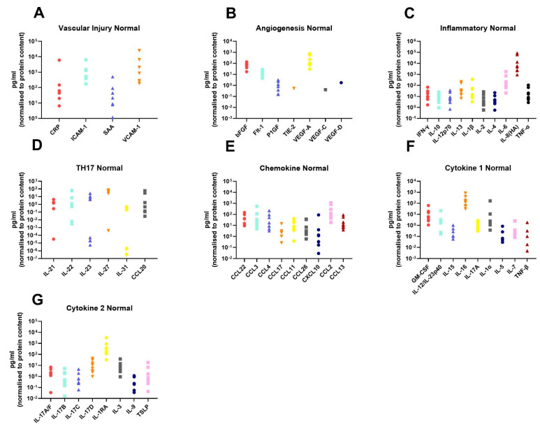
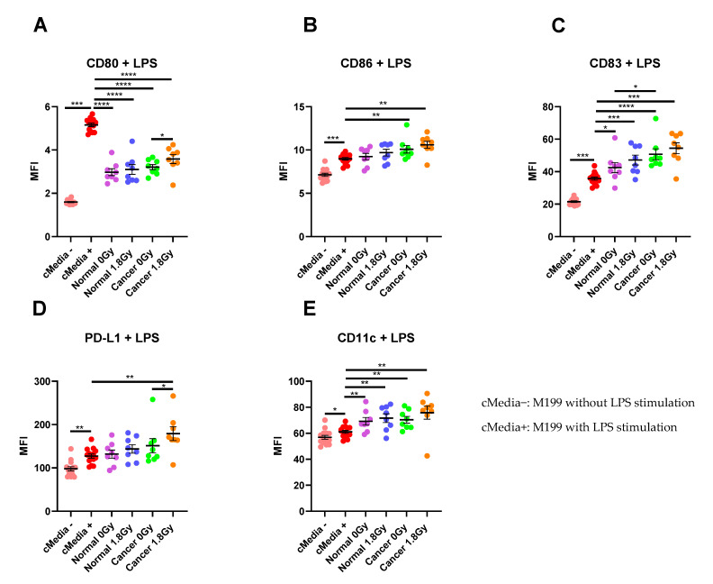
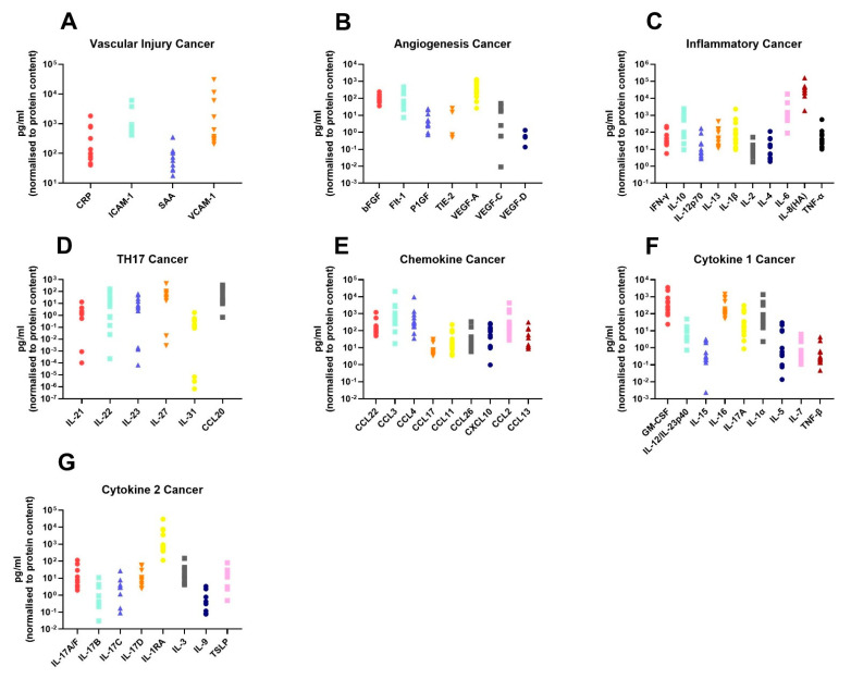
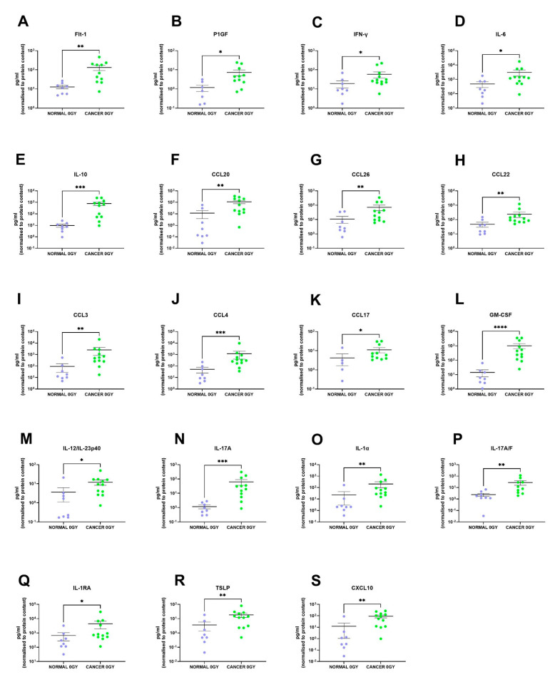
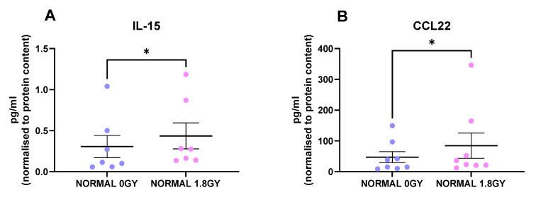
Locally advanced rectal cancer is treated with neoadjuvant-chemoradiotherapy; however, only ~22% of patients achieve a complete response, and resistance mechanisms are poorly understood. The role of inflammation and immune cell biology in this setting is under-investigated. In this study, we profiled the inflammatory protein secretome of normal (non-cancer) ( = 8) and malignant rectal tissue ( = 12) pre- and post-radiation in human ex vivo explant models and examined the influence of these untreated and treated secretomes on dendritic cell biology ( = 8 for cancer and normal). These resultant profiles were correlated with patient clinical characteristics. Nineteen factors were secreted at significantly higher levels from the rectal cancer secretome when compared to the normal rectal secretome; Flt-1, P1GF, IFN-γ, IL-6, IL-10, CCL20, CCL26, CCL22, CCL3, CCL4, CCL17, GM-CSF, IL-12/IL-23p40, IL-17A, IL-1α, IL-17A/F, IL-1RA, TSLP and CXCL10 ( < 0.05). Radiation was found to have differential effects on normal rectal tissue and rectal cancer tissue with increased IL-15 and CCL22 secretion following radiation from normal rectal tissue explants ( < 0.05), while no significant alterations were observed in the irradiated rectal cancer tissue. Interestingly, however, the irradiated rectal cancer secretome induced the most potent effect on dendritic cell maturation via upregulation of CD80 and PD-L1. Patient's visceral fat area correlated with secreted factors including CCL20, suggesting that obesity status may alter the tumour microenvironment (TME). These results suggest that radiation does not have a negative effect on the ability of the rectal cancer TME to induce an immune response. Understanding these responses may unveil potential therapeutic targets to enhance radiation response and mitigate normal tissue injury. Tumour irradiation in this cohort enhances innate immune responses, which may be harnessed to improve patient treatment outcome.
局部晚期直肠癌采用新辅助放化疗进行治疗;然而,只有约22%的患者实现完全缓解,且耐药机制尚不清楚。炎症和免疫细胞生物学在此情况下的作用研究不足。在本研究中,我们在人离体组织模型中分析了正常(非癌)(n = 8)和恶性直肠组织(n = 12)放疗前后的炎性蛋白质分泌组,并研究了这些未处理和处理后的分泌组对树突状细胞生物学的影响(癌组织和正常组织各n = 8)。这些结果与患者的临床特征相关。与正常直肠分泌组相比,直肠癌分泌组中19种因子的分泌水平显著更高;Flt-1、P1GF、IFN-γ、IL-6、IL-10、CCL20、CCL26、CCL22、CCL3、CCL4、CCL#FormatError!、GM-CSF、IL-12/IL-23p40、IL-17A、IL-1α、IL-17A/F、IL-1RA、TSLP和CXCL10(P < 0.05)。发现放疗对正常直肠组织和直肠癌细胞组织有不同影响,正常直肠组织外植体放疗后IL-15和CCL22分泌增加(P < 0.05),而放疗后的直肠癌细胞组织未观察到显著变化。然而,有趣的是,放疗后的直肠癌分泌组通过上调CD80和PD-L1对树突状细胞成熟产生最显著的影响。患者的内脏脂肪面积与包括CCL20在内的分泌因子相关,表明肥胖状态可能会改变肿瘤微环境(TME)。这些结果表明,放疗对直肠癌TME诱导免疫反应的能力没有负面影响。了解这些反应可能会揭示潜在的治疗靶点,以增强放疗反应并减轻正常组织损伤。该队列中的肿瘤放疗增强了先天免疫反应,这可用于改善患者的治疗结果。