Akerele Mercy I, Karakatsanis Nicolas A, Deidda Daniel, Cal-Gonzalez Jacobo, Forsythe Rachael O, Dweck Marc R, Syed Maaz, Newby David E, Aykroyd Robert G, Sourbron Steven, Tsoumpas Charalampos
Biomedical Imaging Science Department, Faculty of Medicine and Health, University of Leeds, UK; Department of Radiology, Weil Cornell Medical College of Cornell University, NY, USA.
Department of Radiology, Weil Cornell Medical College of Cornell University, NY, USA; Biomedical Engineering and Imaging Institute, Icahn School of Medicine at Mount Sinai, NY.
IEEE Trans Radiat Plasma Med Sci. 2020 Jul;4(4):422-432. doi: 10.1109/TRPMS.2020.2980443. Epub 2020 Mar 12.
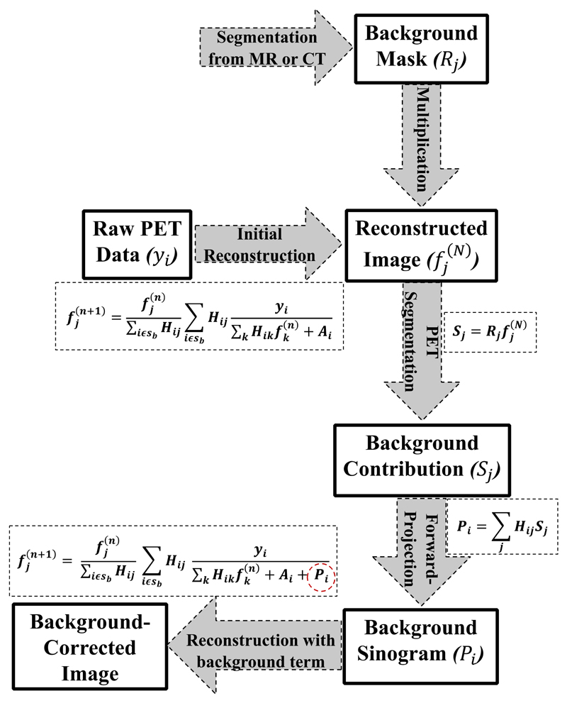
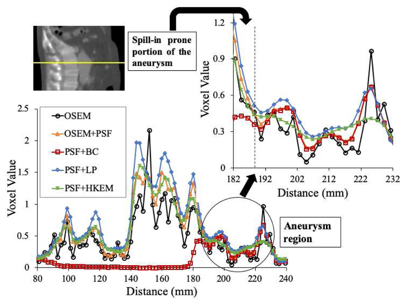
In positron emission tomography (PET) imaging, accurate clinical assessment is often affected by the partial volume effect (PVE) leading to overestimation (spill-in) or underestimation (spill-out) of activity in various small regions. The spill-in correction, in particular, can be very challenging when the target region is close to a hot background region. Therefore, this study evaluates and compares the performance of various recently developed spill-in correction techniques, namely: background correction (BC), local projection (LP), and hybrid kernelized (HKEM) methods. We used a simulated digital phantom and [F]-NaF PET data of three patients with abdominal aortic aneurysms (AAA) acquired with Siemens Biograph mMR™ and mCT™ scanners respectively. Region of Interest (ROI) analysis was performed and the extracted , and target-to-background ratio (TBR) scores were compared. Results showed substantial spill-in effects from hot regions to targeted regions, which are more prominent in small structures. The phantom experiment demonstrated the feasibility of spill-in correction with all methods. For the patient data, large differences in , and scores were observed between the ROIs drawn over the entire aneurysm and ROIs excluding some regions close to the bone. Overall, BC yielded the best performance in spill-in correction in both phantom and patient studies.
在正电子发射断层扫描(PET)成像中,准确的临床评估常常受到部分容积效应(PVE)的影响,导致各种小区域内的活性被高估(溢出效应)或低估(漏出效应)。特别是当目标区域靠近高放射性背景区域时,溢出效应校正可能极具挑战性。因此,本研究评估并比较了各种最近开发的溢出效应校正技术的性能,即:背景校正(BC)、局部投影(LP)和混合核方法(HKEM)。我们使用了一个模拟数字体模以及分别用西门子Biograph mMR™和mCT™扫描仪采集的三名腹主动脉瘤(AAA)患者的[F]-NaF PET数据。进行了感兴趣区域(ROI)分析,并比较了提取的 、 和目标与背景比值(TBR)分数。结果显示从高放射性区域到目标区域存在显著的溢出效应,在小结构中更为突出。体模实验证明了所有方法进行溢出效应校正的可行性。对于患者数据,在整个动脉瘤上绘制的ROI与排除一些靠近骨骼区域的ROI之间,在 、 和 分数上观察到了很大差异。总体而言,在体模和患者研究中,BC在溢出效应校正方面都表现出最佳性能。