Department of Radiation Oncology, Duke University Medical Center, Durham, USA; Department of Pathology, Duke University Medical Center, Durham, USA.
Department of Radiation Oncology, Duke University Medical Center, Durham, USA.
Radiother Oncol. 2021 Apr;157:155-162. doi: 10.1016/j.radonc.2021.01.023. Epub 2021 Feb 3.
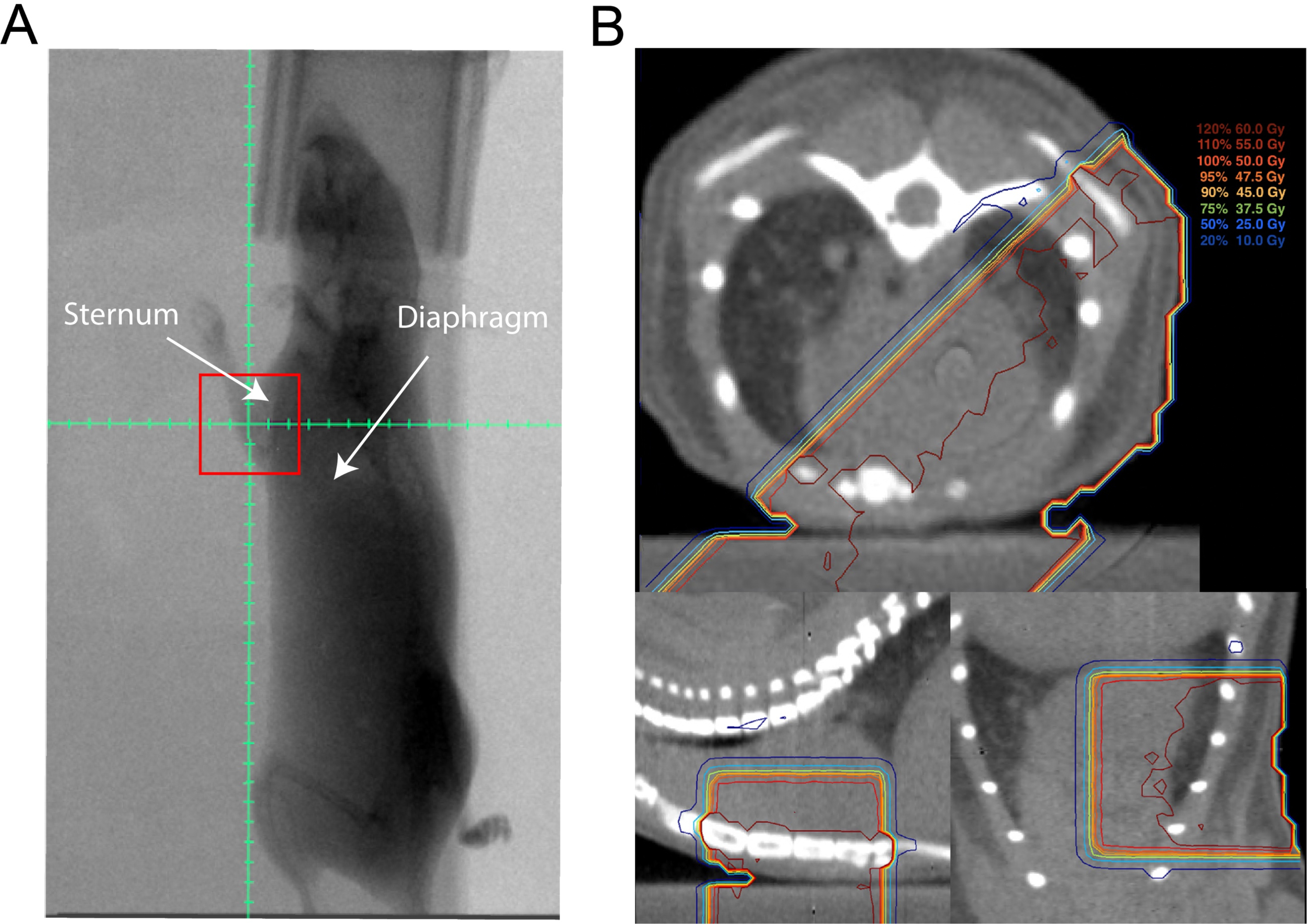
Late cardiac toxicity is a major side effect of radiation therapy (RT) for breast cancer. We developed and characterized a mouse model of radiation-induced heart disease that mimics the dose, fractionation, and beam arrangement of left breast and chest wall RT.
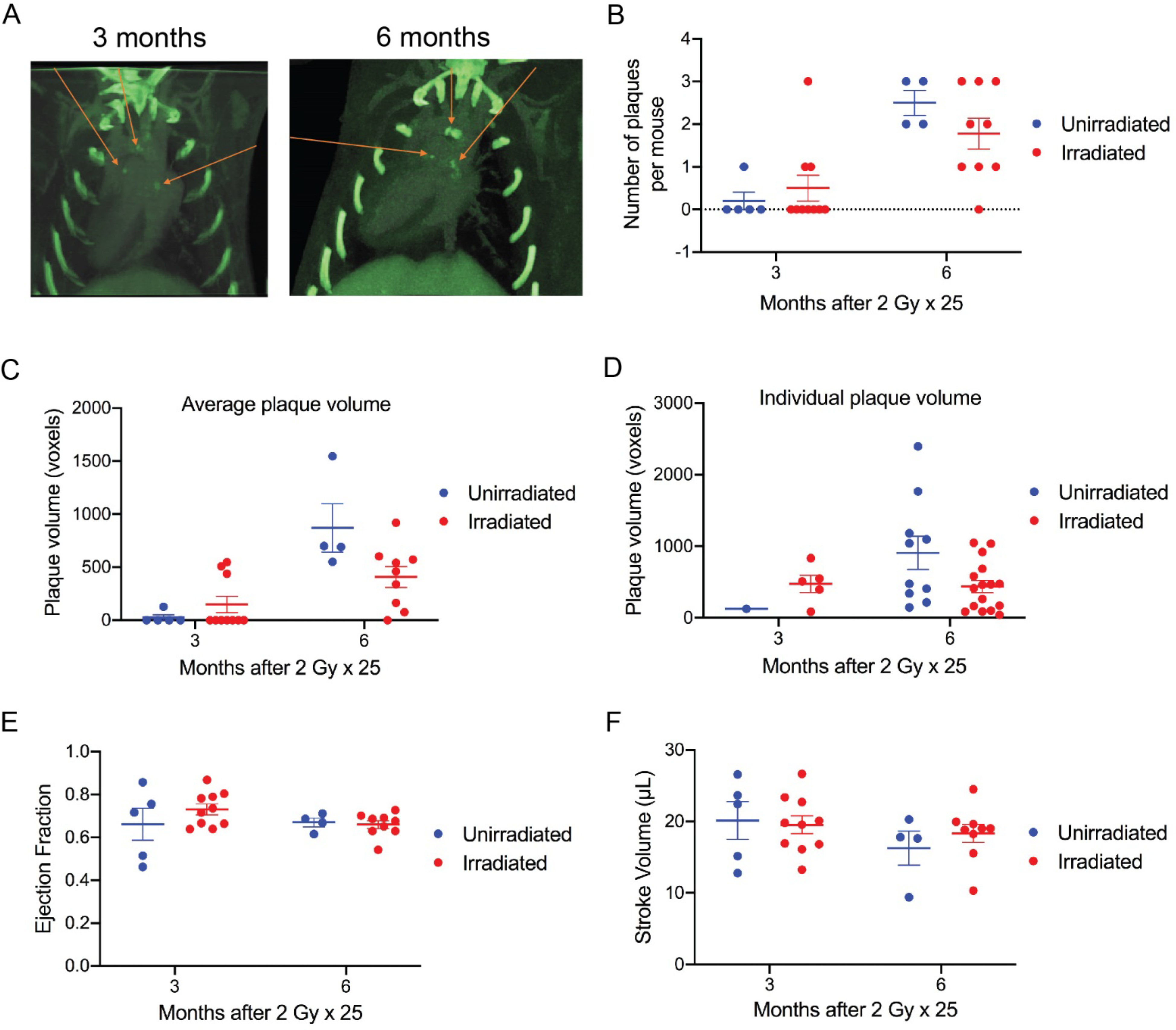
Female wild-type (C57BL6/J) and atherosclerosis-prone apolipoprotein E-deficient (ApoE) mice (on a C57BL/6J background) on regular chow were treated with 2 Gy × 25 fractions of partial-heart irradiation via opposed tangential beams to the left chest wall. The changes in myocardial perfusion and cardiac function of C57BL/6J mice were examined by single-photon emission computed tomography (SPECT) and echocardiography, respectively. In addition to SPECT and echocardiography, the formation of calcified plaques and changes in cardiac function of ApoE mice were examined by dual-energy microCT (DE-CT) and pressure-volume (PV) loop analysis, respectively. The development of myocardial fibrosis was examined by histopathology.
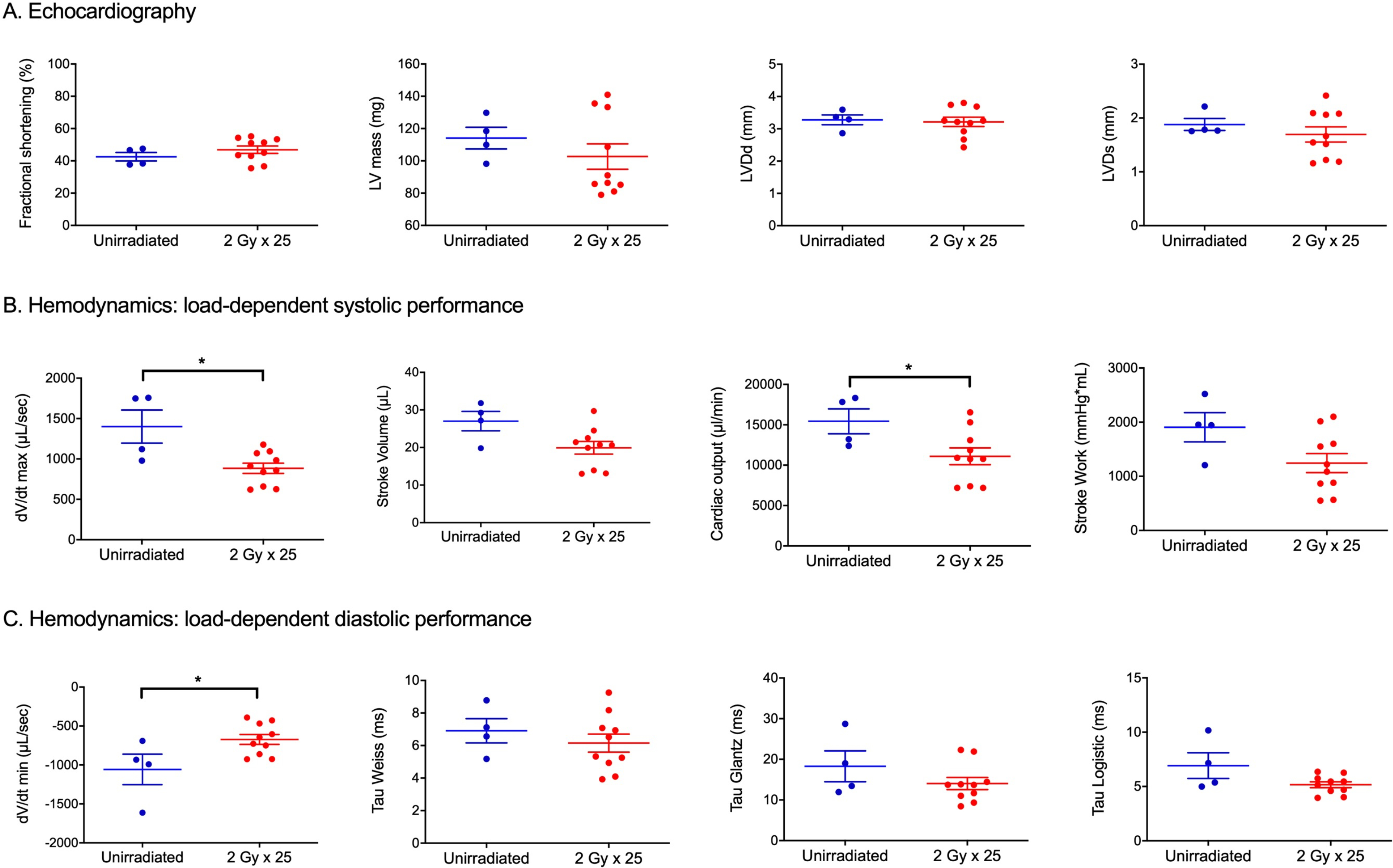
Compared to unirradiated controls, irradiated C57BL/6J mice showed no significant changes by SPECT or echocardiography up to 18 months after 2 Gy × 25 partial-heart irradiation even though irradiated mice exhibited a modest increase in myocardial fibrosis. For ApoE mice, 2 Gy × 25 partial-heart irradiation did not cause significant changes by SPECT, DE-CT, or echocardiography. However, PV loop analysis revealed a significant decrease in load-dependent systolic and diastolic function measures including cardiac output, dV/dtmax and dV/dt min 12 months after RT.
Following clinically relevant doses of partial-heart irradiation in C57BL/6J and ApoE mice, assessment with noninvasive imaging modalities such as echocardiography, SPECT, and DE-CT yielded no evidence of decreased myocardial perfusion and cardiac dysfunction related to RT. However, invasive hemodynamic assessment with PV loop analysis indicated subtle, but significant, changes in cardiac function of irradiated ApoE mice. PV loop analysis may be useful for future preclinical studies of radiation-induced heart disease, especially if subtle changes in cardiac function are expected.
心脏迟发性毒性是乳腺癌放射治疗(RT)的主要副作用。我们开发并鉴定了一种模拟左乳房和胸壁 RT 剂量、分割和射束排列的辐射诱导心脏病小鼠模型。
在普通饲料上,雌性野生型(C57BL6/J)和动脉粥样硬化易感载脂蛋白 E 缺陷(ApoE)小鼠(C57BL/6J 背景)接受 2 Gy×25 次部分心脏照射,采用左胸壁对切射束。通过单光子发射计算机断层扫描(SPECT)和超声心动图分别检测 C57BL/6J 小鼠的心肌灌注和心功能变化。除 SPECT 和超声心动图外,通过双能微 CT(DE-CT)和压力-容积(PV)环分析分别检测 ApoE 小鼠的钙化斑块形成和心功能变化。通过组织病理学检查心肌纤维化的发展情况。
与未照射对照相比,2 Gy×25 次部分心脏照射后 18 个月内,接受照射的 C57BL/6J 小鼠的 SPECT 或超声心动图检查均未见明显变化,尽管照射小鼠的心肌纤维化略有增加。对于 ApoE 小鼠,2 Gy×25 次部分心脏照射后,SPECT、DE-CT 或超声心动图检查均无明显变化。然而,PV 环分析显示,在 RT 后 12 个月时,与负荷相关的收缩期和舒张期功能指标,包括心输出量、dV/dtmax 和 dV/dt min 明显降低。
在 C57BL/6J 和 ApoE 小鼠接受临床相关剂量的部分心脏照射后,非侵入性成像方式如超声心动图、SPECT 和 DE-CT 的评估未发现与 RT 相关的心肌灌注和心功能障碍的证据。然而,PV 环分析显示,照射的 ApoE 小鼠的心脏功能存在微妙但显著的变化。PV 环分析可能对辐射诱导心脏病的未来临床前研究有用,特别是在预计心脏功能会出现细微变化的情况下。