Hamada Nobuyuki, Kawano Ki-Ichiro, Nomura Takaharu, Furukawa Kyoji, Yusoff Farina Mohamad, Maruhashi Tatsuya, Maeda Makoto, Nakashima Ayumu, Higashi Yukihito
Radiation Safety Unit, Biology and Environmental Chemistry Division, Sustainable System Research Laboratory, Central Research Institute of Electric Power Industry (CRIEPI), Tokyo 201-8511, Japan.
Department of Cardiovascular Regeneration and Medicine, Research Institute for Radiation Biology and Medicine, Hiroshima University, Hiroshima 734-8551, Japan.
Cancers (Basel). 2021 Oct 25;13(21):5344. doi: 10.3390/cancers13215344.
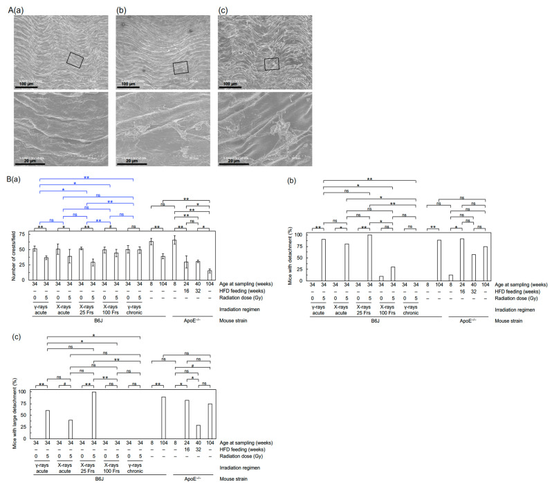
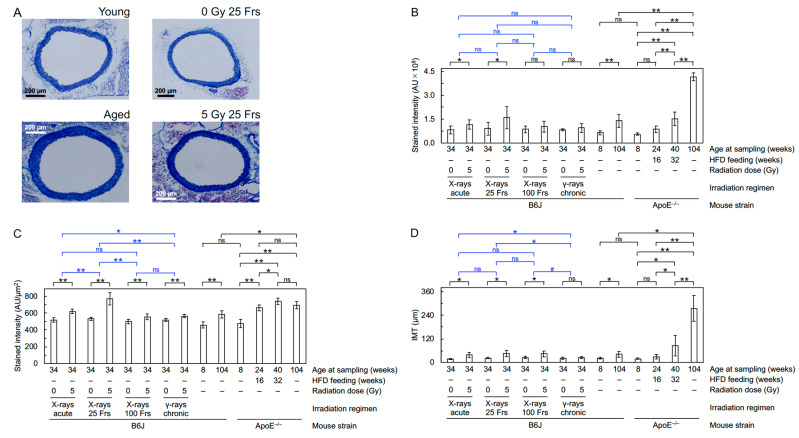
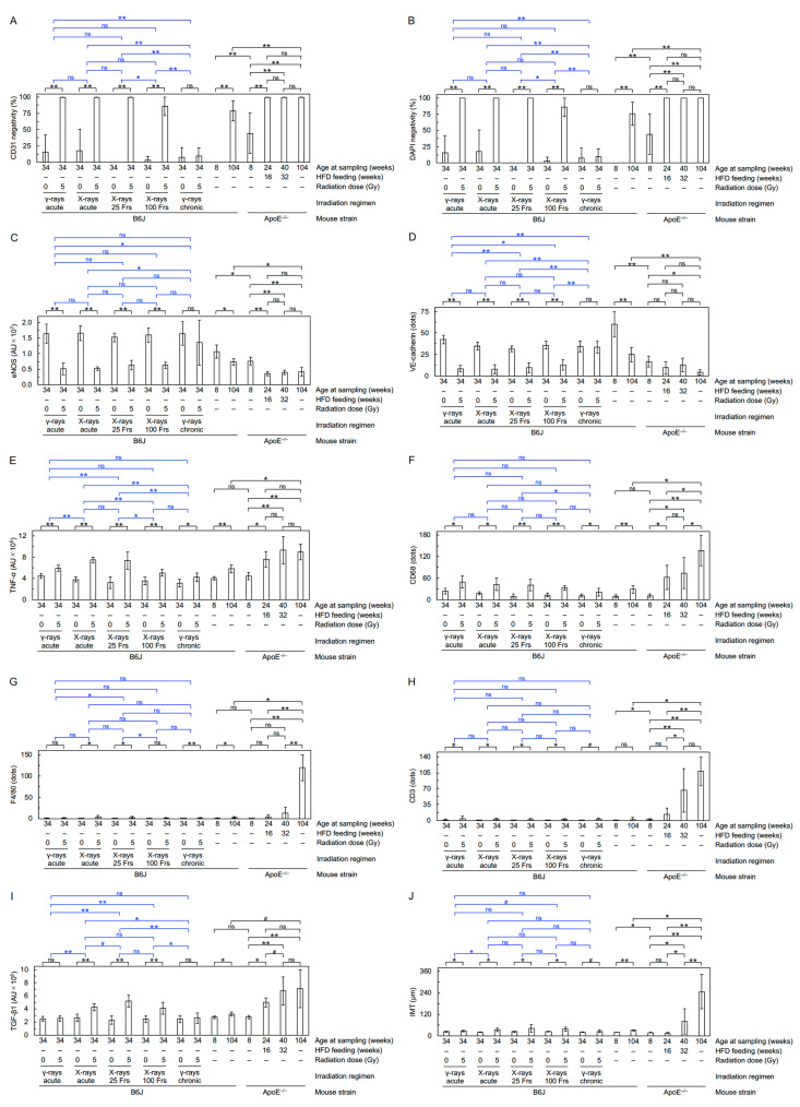
During medical (therapeutic or diagnostic) procedures or in other settings, the circulatory system receives ionizing radiation at various dose rates. Here, we analyzed prelesional changes in the circulatory system of wild-type mice at six months after starting acute, intermittent, or continuous irradiation with 5 Gy of photons. Independent of irradiation regimens, irradiation had little impact on left ventricular function, heart weight, and kidney weight. In the aorta, a single acute exposure delivered in 10 minutes led to structural disorganizations and detachment of the aortic endothelium, and intima-media thickening. These morphological changes were accompanied by increases in markers for profibrosis (TGF-β1), fibrosis (collagen fibers), proinflammation (TNF-α), and macrophages (F4/80 and CD68), with concurrent decreases in markers for cell adhesion (CD31 and VE-cadherin) and vascular functionality (eNOS) in the aortic endothelium. Compared with acute exposure, the magnitude of such aortic changes was overall greater when the same dose was delivered in 25 fractions spread over 6 weeks, smaller in 100 fractions over 5 months, and much smaller in chronic exposure over 5 months. These findings suggest that dose protraction alters vascular damage in the aorta, but in a way that is not a simple function of dose rate.
在医疗(治疗或诊断)程序期间或在其他情况下,循环系统会以不同的剂量率接受电离辐射。在此,我们分析了野生型小鼠在开始用5 Gy光子进行急性、间歇性或连续照射六个月后的循环系统损伤前变化。与照射方案无关,照射对左心室功能、心脏重量和肾脏重量影响很小。在主动脉中,10分钟内进行的单次急性照射导致主动脉内皮结构紊乱和脱离,以及内膜中层增厚。这些形态学变化伴随着促纤维化标志物(TGF-β1)、纤维化(胶原纤维)、促炎标志物(TNF-α)和巨噬细胞(F4/80和CD68)的增加,同时主动脉内皮细胞黏附标志物(CD31和VE-钙黏蛋白)和血管功能标志物(eNOS)减少。与急性照射相比,当相同剂量在6周内分25次给予时,主动脉这种变化的程度总体上更大;在5个月内分100次给予时较小;在5个月的慢性照射时则小得多。这些发现表明,剂量分割会改变主动脉的血管损伤,但并非简单地取决于剂量率。