Department of Radiology & Nuclear Medicine, Amsterdam Neuroscience, Vrije Universiteit Amsterdam, Amsterdam UMC, Amsterdam, The Netherlands.
Alzheimer Center Amsterdam, Department of Neurology, Amsterdam Neuroscience, Vrije Universiteit Amsterdam, Amsterdam UMC, Amsterdam, The Netherlands.
Alzheimers Res Ther. 2021 Feb 5;13(1):35. doi: 10.1186/s13195-021-00772-0.
The mechanism of synaptic loss in Alzheimer's disease is poorly understood and may be associated with tau pathology. In this combined positron emission tomography (PET) and magnetoencephalography (MEG) study, we aimed to investigate spatial associations between regional tau pathology ([F]flortaucipir PET), synaptic density (synaptic vesicle 2A [C]UCB-J PET) and synaptic function (MEG) in Alzheimer's disease.
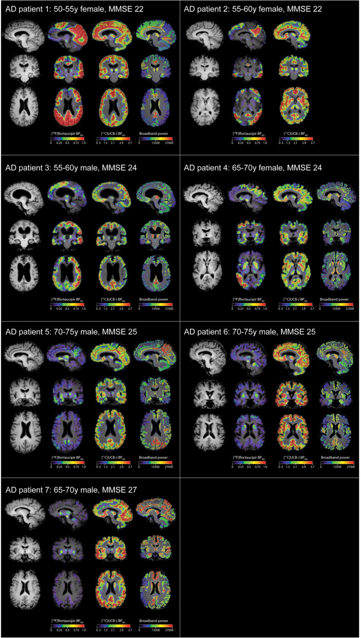
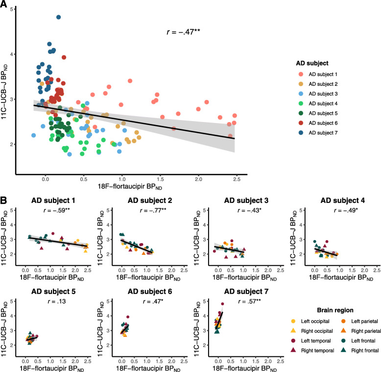
Seven amyloid-positive Alzheimer's disease subjects from the Amsterdam Dementia Cohort underwent dynamic 130-min [F]flortaucipir PET, dynamic 60-min [C]UCB-J PET with arterial sampling and 2 × 5-min resting-state MEG measurement. [F]flortaucipir- and [C]UCB-J-specific binding (binding potential, BP) and MEG spectral measures (relative delta, theta and alpha power; broadband power; and peak frequency) were assessed in cortical brain regions of interest. Associations between regional [F]flortaucipir BP, [C]UCB-J BP and MEG spectral measures were assessed using Spearman correlations and generalized estimating equation models.
Across subjects, higher regional [F]flortaucipir uptake was associated with lower [C]UCB-J uptake. Within subjects, the association between [C]UCB-J and [F]flortaucipir depended on within-subject neocortical tau load; negative associations were observed when neocortical tau load was high, gradually changing into opposite patterns with decreasing neocortical tau burden. Both higher [F]flortaucipir and lower [C]UCB-J uptake were associated with altered synaptic function, indicative of slowing of oscillatory activity, most pronounced in the occipital lobe.
These results indicate that in Alzheimer's disease, tau pathology is closely associated with reduced synaptic density and synaptic dysfunction.
阿尔茨海默病中突触丧失的机制尚未完全阐明,可能与 tau 病理学有关。在这项正电子发射断层扫描(PET)和脑磁图(MEG)联合研究中,我们旨在研究阿尔茨海默病患者中局部 tau 病理学([F]flortaucipir PET)、突触密度(突触小泡 2A [C]UCB-J PET)和突触功能(MEG)之间的空间关联。
来自阿姆斯特丹痴呆队列的 7 名淀粉样蛋白阳性阿尔茨海默病患者接受了 130 分钟的动态 [F]flortaucipir PET、60 分钟的动态 [C]UCB-J PET 伴动脉采样和 2×5 分钟静息状态 MEG 测量。在皮质脑感兴趣区评估 [F]flortaucipir 和 [C]UCB-J 特异性结合(结合潜能,BP)和 MEG 光谱测量(相对 delta、theta 和 alpha 功率;宽带功率;和峰值频率)。使用 Spearman 相关和广义估计方程模型评估局部 [F]flortaucipir BP、[C]UCB-J BP 和 MEG 光谱测量之间的相关性。
在所有患者中,较高的区域 [F]flortaucipir 摄取与较低的 [C]UCB-J 摄取相关。在个体内,[C]UCB-J 和 [F]flortaucipir 之间的关联取决于个体内的新皮质 tau 负荷;当新皮质 tau 负荷较高时,观察到负相关,随着新皮质 tau 负担的逐渐降低,逐渐呈现相反的模式。较高的 [F]flortaucipir 和较低的 [C]UCB-J 摄取均与突触功能改变相关,表明振荡活动减慢,在枕叶中最为明显。
这些结果表明,在阿尔茨海默病中,tau 病理学与突触密度降低和突触功能障碍密切相关。