Department of Biophysics, Bose Institute, Kolkata 700 054, India.
School of Biological Sciences, Nanyang Technological University, Singapore 637541, Singapore.
Int J Biol Macromol. 2021 Apr 1;175:131-139. doi: 10.1016/j.ijbiomac.2021.02.011. Epub 2021 Feb 4.
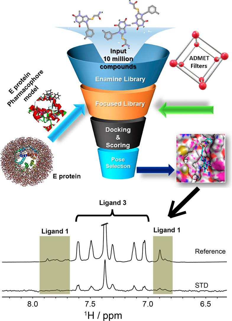
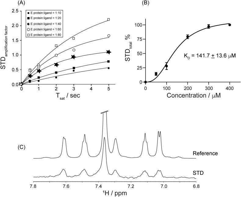
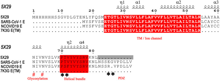
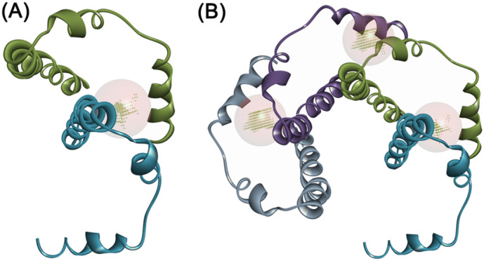
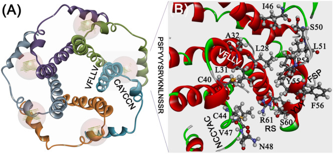
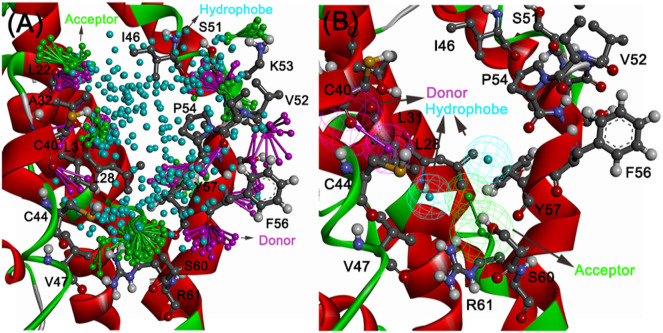
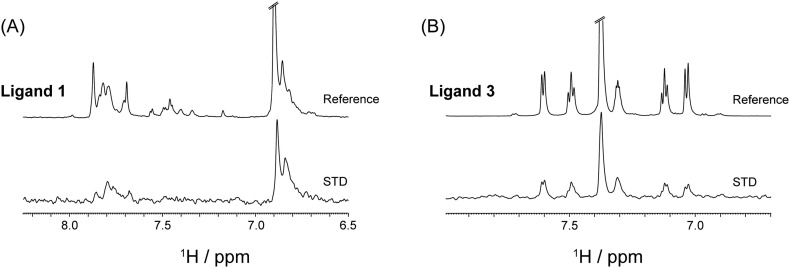
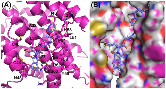
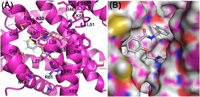
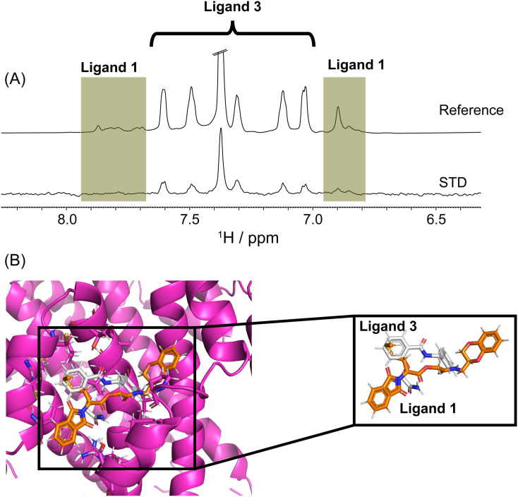
One of the most crucial characteristic traits of Envelope (E) proteins in the severe acute respiratory syndrome SARS-CoV-1 and NCOVID19 viruses is their membrane-associated oligomerization led ion channel activity, virion assembly, and replication. NMR spectroscopic structural studies of envelope proteins from both the SARS CoV-1/2 reveal that this protein assembles into a homopentamer. Proof of concept studies via truncation mutants on either transmembrane (VFLLV), glycosylation motif (CACCN), hydrophobic helical bundle (PVYVY) as well as replacing C-terminal "DLLV" segments or point mutants such as S68, E69 residues with cysteine have significantly reduced viral titers of SARS-CoV-1. In this present study, we have first developed SARS-2 E protein homology model based on the pentamer coordinates of SARS-CoV-1 E protein (86.4% structural identity) with good stereochemical quality. Next, we focused on the glycosylation motif and hydrophobic helical bundle regions of E protein shown to be important for viral replication. A four feature (4F) model comprising of an acceptor targeting S60 hydroxyl group, a donor feature anchoring the C40 residue, and two hydrophobic features anchoring the V47 L28, L31, Y55, and P51 residues formed the protein based pharmacophore model targeting the glycosylation motif and helical bundle of E protein. Database screening with this 4F protein pharmacophore, ADMET property filtering on enamine small molecule discovery collection yielded a focused library of ~7000 hits. Further molecular docking and visual inspection of docked pose interactions at the above mention V47 L28, L31, Y55, P51, S60, C40 residues led to the identification of 10 best hits. Our STD NMR binding assay results demonstrate that the ligand 3, 2-(2-amino-2-oxo-ethoxy)-N-benzyl-benzamide, binds to NCOVID19 E protein with a binding affinity (K) of 141.7 ± 13.6 μM. Furthermore, the ligand 3 also showed binding to C-terminal peptide (NR25) as evidenced with the STD spectrums of wild type E protein would serve to confirm the involvement of C-terminal helical bundle as envisaged in this study.
严重急性呼吸系统综合症冠状病毒 SARS-CoV-1 和新型冠状病毒 NCOVID19 的包膜(E)蛋白的最重要特征之一是其与膜相关的寡聚化导致的离子通道活性、病毒体组装和复制。对来自 SARS CoV-1/2 的包膜蛋白的 NMR 光谱结构研究表明,这种蛋白质组装成同源五聚体。通过对跨膜(VFLLV)、糖基化模体(CACCN)、疏水性螺旋束(PVYVY)的截断突变体,以及用半胱氨酸替换 C 末端“DLLV”片段或点突变(如 S68、E69 残基)进行的概念验证研究,显著降低了 SARS-CoV-1 的病毒滴度。在本研究中,我们首先基于 SARS-CoV-1 E 蛋白的五聚体坐标(86.4%的结构同一性),开发了 SARS-2 E 蛋白同源模型,具有良好的立体化学质量。接下来,我们集中研究了对病毒复制很重要的 E 蛋白的糖基化模体和疏水性螺旋束区域。一个由四个特征(4F)组成的模型,包括一个靶向 S60 羟基的受体、一个锚定 C40 残基的供体特征,以及两个锚定 V47、L28、L31、Y55 和 P51 残基的疏水性特征,形成了针对 E 蛋白糖基化模体和螺旋束的基于配体的药效团模型。使用该 4F 蛋白药效团模型对数据库进行筛选,并对 enamine 小分子发现库的 ADMET 性质进行过滤,得到了约 7000 个命中化合物的聚焦文库。进一步的分子对接和对接构象在上述 V47、L28、L31、Y55、P51、S60、C40 残基的相互作用的可视化检查,导致鉴定出 10 个最佳配体。我们的 STD NMR 结合测定结果表明,配体 3, 2-(2-氨基-2-氧代-乙氧基)-N-苄基-苯甲酰胺,与 NCOVID19 E 蛋白的结合亲和力(K)为 141.7±13.6μM。此外,配体 3 还与 C 末端肽(NR25)结合,这一点从野生型 E 蛋白的 STD 谱中可以得到证实,这将证实本研究中所设想的 C 末端螺旋束的参与。