• 文献检索
  • 文档翻译
  • 深度研究
  • 学术资讯
  • Suppr Zotero 插件Zotero 插件
  • 邀请有礼
  • 套餐&价格
  • 历史记录
应用&插件
Suppr Zotero 插件Zotero 插件浏览器插件Mac 客户端Windows 客户端微信小程序
定价
高级版会员购买积分包购买API积分包
服务
文献检索文档翻译深度研究API 文档MCP 服务
关于我们
关于 Suppr公司介绍联系我们用户协议隐私条款
关注我们

Suppr 超能文献

核心技术专利:CN118964589B侵权必究
粤ICP备2023148730 号-1Suppr @ 2026

文献检索

告别复杂PubMed语法,用中文像聊天一样搜索,搜遍4000万医学文献。AI智能推荐,让科研检索更轻松。

立即免费搜索

文件翻译

保留排版,准确专业,支持PDF/Word/PPT等文件格式,支持 12+语言互译。

免费翻译文档

深度研究

AI帮你快速写综述,25分钟生成高质量综述,智能提取关键信息,辅助科研写作。

立即免费体验

日本窄带成像专家组分类法对结直肠病变的诊断能力:蓝激光成像放大内镜与窄带成像的比较

Diagnostic ability of Japan Narrow-Band Imaging Expert Team classification for colorectal lesions by magnifying endoscopy with blue laser imaging versus narrow-band imaging.

作者信息

Ito Renma, Ikematsu Hiroaki, Murano Tatsuro, Shinmura Kensuke, Kojima Motohiro, Kumahara Kana, Furue Yasuaki, Sunakawa Hironori, Minamide Tatsunori, Sato Daiki, Yamamoto Yoichi, Takashima Kenji, Yoda Yusuke, Hori Keisuke, Yano Tomonori

机构信息

Department of Gastroenterology and Endoscopy.

Division of Pathology, National Cancer Center Hospital East.

出版信息

Endosc Int Open. 2021 Feb;9(2):E271-E277. doi: 10.1055/a-1324-3083. Epub 2021 Feb 3.

DOI:10.1055/a-1324-3083
PMID:33553592
原文链接:https://pmc.ncbi.nlm.nih.gov/articles/PMC7857969/
Abstract

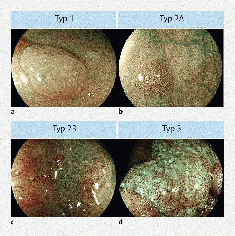
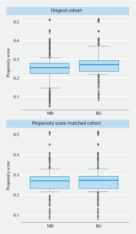
The Japan Narrow-band imaging (NBI) Expert Team (JNET) classification was proposed for evaluating colorectal lesions. However, it remains unknown whether the JNET classification can be applied to magnifying endoscopy with image-enhanced endoscopies other than NBI. This study aimed to compare the diagnostic ability of JNET classification by magnifying endoscopy with blue laser imaging (ME-BLI) and with ME-NBI. We retrospectively assessed consecutive patients diagnosed per the JNET classification by ME-BLI (BLI group) or ME-NBI (NBI group) between March 2014 and June 2017. We compared the diagnostic value of JNET classification between the groups with one-to-one propensity score matching. Four hundred and seventy-one propensity score-matched pairs of lesions were analyzed. In the BLI and NBI groups, the overall diagnostic accuracies were 92.1 % and 91.7 %, respectively, and those for differentiating between neoplastic and non-neoplastic polyps were 96.6 % and 96.8 %, respectively. The positive predictive value by each JNET classification in BLI vs. NBI group was 90.6 % vs. 96.2 % in Type 1, 94.3 % vs. 94.6 % in Type 2A, 57.7 % vs. 42.3 % in Type 2B, and 100 % vs. 91.7 % in Type 3. The negative predictive value was 97.0 % vs. 96.9 % in Type 1, 88.1 % vs. 82.8 % in Type 2A, 98.0 % vs. 98.2 % in Type 2B, and 98.5 % vs. 98.7 % in Type 3. No statistical difference in the diagnostic results was found between the groups. The diagnostic ability of the JNET classification by ME-BLI and ME-NBI was comparable, with the former also applicable for diagnosis of colorectal lesions.

摘要

日本窄带成像(NBI)专家组(JNET)分类法被提出用于评估结直肠病变。然而,JNET分类法是否可应用于除NBI之外的图像增强型内镜的放大内镜检查仍不清楚。本研究旨在比较通过放大内镜联合蓝光成像(ME-BLI)和联合ME-NBI的JNET分类法的诊断能力。我们回顾性评估了2014年3月至2017年6月期间根据ME-BLI(BLI组)或ME-NBI(NBI组)按照JNET分类法诊断的连续患者。我们采用一对一倾向评分匹配比较了两组之间JNET分类法的诊断价值。分析了471对倾向评分匹配的病变。在BLI组和NBI组中,总体诊断准确率分别为92.1%和91.7%,区分肿瘤性息肉和非肿瘤性息肉的准确率分别为96.6%和96.8%。BLI组与NBI组中各JNET分类的阳性预测值在1型中分别为90.6%和96.2%,2A型中分别为94.3%和94.6%,2B型中分别为57.7%和42.3%,3型中分别为100%和91.7%。阴性预测值在1型中分别为97.0%和96.9%,2A型中分别为88.1%和82.8%,2B型中分别为98.0%和98.2%,3型中分别为98.5%和98.7%。两组之间的诊断结果未发现统计学差异。ME-BLI和ME-NBI的JNET分类法的诊断能力相当,前者也适用于结直肠病变的诊断。

https://cdn.ncbi.nlm.nih.gov/pmc/blobs/5104/7857969/39ecf4521a31/10-1055-a-1324-3083-i2114ei2.jpg
https://cdn.ncbi.nlm.nih.gov/pmc/blobs/5104/7857969/ea64dc459ad9/10-1055-a-1324-3083-i2114ei1.jpg
https://cdn.ncbi.nlm.nih.gov/pmc/blobs/5104/7857969/39ecf4521a31/10-1055-a-1324-3083-i2114ei2.jpg
https://cdn.ncbi.nlm.nih.gov/pmc/blobs/5104/7857969/ea64dc459ad9/10-1055-a-1324-3083-i2114ei1.jpg
https://cdn.ncbi.nlm.nih.gov/pmc/blobs/5104/7857969/39ecf4521a31/10-1055-a-1324-3083-i2114ei2.jpg

相似文献

1
Diagnostic ability of Japan Narrow-Band Imaging Expert Team classification for colorectal lesions by magnifying endoscopy with blue laser imaging versus narrow-band imaging.日本窄带成像专家组分类法对结直肠病变的诊断能力:蓝激光成像放大内镜与窄带成像的比较
Endosc Int Open. 2021 Feb;9(2):E271-E277. doi: 10.1055/a-1324-3083. Epub 2021 Feb 3.
2
Magnifying Narrow Band Imaging (NBI) for the Diagnosis of Localized Colorectal Lesions Using the Japan NBI Expert Team (JNET) Classification.使用日本窄带成像专家团队(JNET)分类法的放大窄带成像(NBI)用于诊断局限性结直肠病变
Oncology. 2017;93 Suppl 1:49-54. doi: 10.1159/000481230. Epub 2017 Dec 20.
3
Diagnostic performance of narrow-band imaging international colorectal endoscopic and Japanese narrow-band imaging expert team classification systems for colorectal cancer and precancerous lesions.窄带成像国际结直肠内镜和日本窄带成像专家组分类系统对结直肠癌及癌前病变的诊断性能
World J Gastrointest Oncol. 2021 Jan 15;13(1):58-68. doi: 10.4251/wjgo.v13.i1.58.
4
Diagnostic efficacy of the Japan NBI Expert Team classification with dual-focus magnification for colorectal tumors.日本 NBI 专家小组分类法(双焦距放大)对结直肠肿瘤的诊断效能。
Surg Endosc. 2022 Jul;36(7):5032-5040. doi: 10.1007/s00464-021-08863-7. Epub 2021 Nov 29.
5
Comparison of the diagnostic performance of NBI, Laser-BLI and LED-BLI: a randomized controlled noninferiority trial.窄带成像、激光-蓝激光成像和白光成像诊断性能的比较:一项随机对照非劣效性试验。
Surg Endosc. 2022 Oct;36(10):7577-7587. doi: 10.1007/s00464-022-09197-8. Epub 2022 Apr 11.
6
Diagnostic yield of the Japan NBI Expert Team (JNET) classification for endoscopic diagnosis of superficial colorectal neoplasms in a large-scale clinical practice database.日本 NBI 专家团队(JNET)分类法在大规模临床实践数据库中用于内镜诊断浅表结直肠肿瘤的诊断率。
United European Gastroenterol J. 2019 Aug;7(7):914-923. doi: 10.1177/2050640619845987. Epub 2019 Apr 26.
7
Effect of educational lecture on the diagnostic accuracy of Japan NBI Expert Team classification for colorectal lesions.教育讲座对日本 NBI 专家分类法诊断结直肠病变准确性的影响。
BMC Gastroenterol. 2021 Mar 5;21(1):110. doi: 10.1186/s12876-021-01676-x.
8
Usefulness of the Japan narrow-band imaging expert team classification system for the diagnosis of sessile serrated lesion with dysplasia/carcinoma.日本窄带成像专家分类系统对无蒂锯齿状病变伴异型增生/癌的诊断价值。
Surg Endosc. 2021 Aug;35(8):4528-4538. doi: 10.1007/s00464-020-07967-w. Epub 2020 Sep 9.
9
Additional chromoendoscopy for colorectal lesions initially diagnosed with low confidence by magnifying narrow-band imaging: Can it improve diagnostic accuracy? 对放大窄带成像初步诊断信心较低的结直肠病变行额外 chromoendoscopy:是否能提高诊断准确性?
Dig Endosc. 2018 Apr;30 Suppl 1:45-50. doi: 10.1111/den.13055.
10
An International Study on the Diagnostic Accuracy of the Japan Narrow-Band Imaging Expert Team Classification for Colorectal Polyps Observed with Blue Laser Imaging.国际研究:蓝色激光成像观察下日本窄带成像专家团队分类法对结直肠息肉的诊断准确性。
Digestion. 2020;101(3):339-346. doi: 10.1159/000499856. Epub 2019 Apr 12.

引用本文的文献

1
Imaging colonic polyps in 2024.2024 年的结肠息肉成像。
Indian J Gastroenterol. 2024 Oct;43(5):954-965. doi: 10.1007/s12664-024-01679-y. Epub 2024 Sep 30.
2
Use of artificial intelligence in the management of T1 colorectal cancer: a new tool in the arsenal or is deep learning out of its depth?人工智能在T1期结直肠癌管理中的应用:武器库中的新工具还是深度学习力有不逮?
Clin Endosc. 2024 Jan;57(1):24-35. doi: 10.5946/ce.2023.036. Epub 2023 Sep 25.
3
Image enhanced colonoscopy: updates and prospects-a review.图像增强结肠镜检查:最新进展与前景——综述

本文引用的文献

1
Blue Laser Imaging, Blue Light Imaging, and Linked Color Imaging for the Detection and Characterization of Colorectal Tumors.蓝激光成像、蓝光成像和链接彩色成像在结直肠肿瘤检测和特征分析中的应用。
Gut Liver. 2019 Mar 15;13(2):140-148. doi: 10.5009/gnl18276.
2
Validation study for development of the Japan NBI Expert Team classification of colorectal lesions.日本 NBI 专家小组分类法用于结直肠病变的开发验证研究。
Dig Endosc. 2018 Sep;30(5):642-651. doi: 10.1111/den.13065. Epub 2018 Jun 26.
3
Magnifying Narrow Band Imaging (NBI) for the Diagnosis of Localized Colorectal Lesions Using the Japan NBI Expert Team (JNET) Classification.
Transl Gastroenterol Hepatol. 2023 Jul 25;8:26. doi: 10.21037/tgh-23-17. eCollection 2023.
4
Mucosal imaging in colon polyps: New advances and what the future may hold.结肠息肉的黏膜成像:新进展和未来可能的发展方向。
World J Gastroenterol. 2022 Dec 21;28(47):6632-6661. doi: 10.3748/wjg.v28.i47.6632.
5
Brown slits for colorectal adenoma crypts on conventional magnifying endoscopy with narrow band imaging using the X1 system.常规放大内镜窄带成像 X1 系统下结肠直肠腺瘤隐窝的棕色裂隙。
World J Gastroenterol. 2022 Jun 28;28(24):2748-2757. doi: 10.3748/wjg.v28.i24.2748.
6
The Efficacy of Tumor Characterization for Colorectal Lesions with Blue Light Imaging of a Compact Light-Emitting Diode Endoscopic System Compared to a Laser Endoscopic System: A Pilot Study.与激光内镜系统相比,紧凑型发光二极管内镜系统蓝光成像对结直肠病变的肿瘤特征识别效能:一项初步研究
Gastroenterol Res Pract. 2022 Apr 12;2022:9998280. doi: 10.1155/2022/9998280. eCollection 2022.
使用日本窄带成像专家团队(JNET)分类法的放大窄带成像(NBI)用于诊断局限性结直肠病变
Oncology. 2017;93 Suppl 1:49-54. doi: 10.1159/000481230. Epub 2017 Dec 20.
4
BASIC (BLI Adenoma Serrated International Classification) classification for colorectal polyp characterization with blue light imaging.基于蓝光成像的结直肠息肉特征的 BASIC(BLI 锯齿状腺瘤国际分类)分类。
Endoscopy. 2018 Mar;50(3):211-220. doi: 10.1055/s-0043-121570. Epub 2017 Oct 24.
5
Diagnostic performance of Japan NBI Expert Team classification for differentiation among noninvasive, superficially invasive, and deeply invasive colorectal neoplasia.日本 NBI 专家分类法对非侵袭性、表浅性和深部侵袭性结直肠肿瘤的鉴别诊断性能。
Gastrointest Endosc. 2017 Oct;86(4):700-709. doi: 10.1016/j.gie.2017.02.018. Epub 2017 Feb 28.
6
Clinical impact and characteristics of the narrow-band imaging magnifying endoscopic classification of colorectal tumors proposed by the Japan NBI Expert Team.日本窄带成像放大内镜专家组提出的结直肠肿瘤窄带成像放大内镜分类的临床影响和特征。
Gastrointest Endosc. 2017 Apr;85(4):816-821. doi: 10.1016/j.gie.2016.07.035. Epub 2016 Jul 25.
7
Narrow-band imaging (NBI) magnifying endoscopic classification of colorectal tumors proposed by the Japan NBI Expert Team.日本窄带成像(NBI)专家组提出的结直肠肿瘤窄带成像(NBI)放大内镜分类。
Dig Endosc. 2016 Jul;28(5):526-33. doi: 10.1111/den.12644. Epub 2016 Apr 20.
8
Effect of novel bright image enhanced endoscopy using blue laser imaging (BLI).新型蓝光成像(BLI)增强内镜明亮图像的效果。
Endosc Int Open. 2014 Dec;2(4):E212-9. doi: 10.1055/s-0034-1390707. Epub 2014 Oct 24.
9
ASGE Technology Committee systematic review and meta-analysis assessing the ASGE PIVI thresholds for adopting real-time endoscopic assessment of the histology of diminutive colorectal polyps.ASGE 技术委员会的系统评价和荟萃分析评估了 ASGE PIVI 阈值,以实时评估微小结直肠息肉的组织学。
Gastrointest Endosc. 2015 Mar;81(3):502.e1-502.e16. doi: 10.1016/j.gie.2014.12.022. Epub 2015 Jan 16.
10
Ability of a novel blue laser imaging system for the diagnosis of colorectal polyps.一种新型蓝色激光成像系统诊断大肠息肉的能力。
Dig Endosc. 2014 Mar;26(2):250-8. doi: 10.1111/den.12127. Epub 2013 Jun 3.