• 文献检索
  • 文档翻译
  • 深度研究
  • 学术资讯
  • Suppr Zotero 插件Zotero 插件
  • 邀请有礼
  • 套餐&价格
  • 历史记录
应用&插件
Suppr Zotero 插件Zotero 插件浏览器插件Mac 客户端Windows 客户端微信小程序
定价
高级版会员购买积分包购买API积分包
服务
文献检索文档翻译深度研究API 文档MCP 服务
关于我们
关于 Suppr公司介绍联系我们用户协议隐私条款
关注我们

Suppr 超能文献

核心技术专利:CN118964589B侵权必究
粤ICP备2023148730 号-1Suppr @ 2026

文献检索

告别复杂PubMed语法,用中文像聊天一样搜索,搜遍4000万医学文献。AI智能推荐,让科研检索更轻松。

立即免费搜索

文件翻译

保留排版,准确专业,支持PDF/Word/PPT等文件格式,支持 12+语言互译。

免费翻译文档

深度研究

AI帮你快速写综述,25分钟生成高质量综述,智能提取关键信息,辅助科研写作。

立即免费体验

骨巨细胞瘤:从眶内肿物到骨内肿物的行为变化

Giant Cell Tumor: Changing Behavior from Intraorbital to Intraosseous Mass.

作者信息

Rajabi Mohammad Taher, Poursayed Lazarjani Seyedeh Zahra, Mohammadi S Saeed, Veshagh Mohammad, Hosseinzadeh Farideh, Rafizadeh Seyed Mohsen, Amoli Fahimeh Asadi, Hosseini Simindokht

机构信息

Eye Research Center, Farabi Eye Hospital, Tehran University of Medical Sciences, Tehran, Iran.

Eye Research Center, Labbafinejad Eye Hospital, Shahid Beheshti University of Medical Sciences, Tehran, Iran.

出版信息

J Curr Ophthalmol. 2020 Dec 12;32(4):414-416. doi: 10.4103/JOCO.JOCO_63_20. eCollection 2020 Oct-Dec.

DOI:10.4103/JOCO.JOCO_63_20
PMID:33553846
原文链接:https://pmc.ncbi.nlm.nih.gov/articles/PMC7861103/
Abstract

PURPOSE

To present a patient with giant cell tumor (GCT) of the orbit by changing behavior from an intraorbital mass to an intraosseous tumor.

METHODS

A 16-year-old boy presented with pain, swelling, erythematous of the left upper and lower eyelids, proptosis, and diplopia. Ophthalmic examination revealed chemosis, conjunctival injection, limited elevation, depression as well as abduction in the left eye.

RESULTS

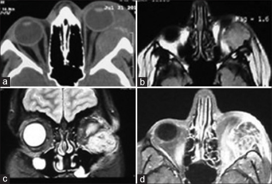
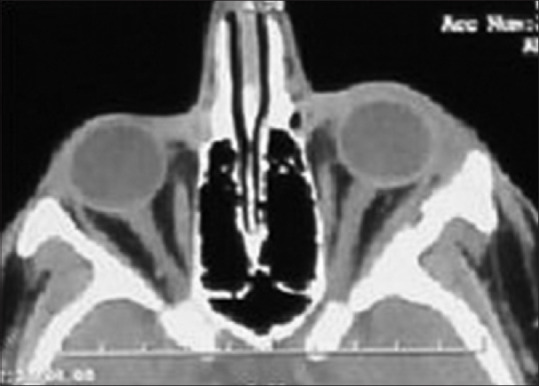
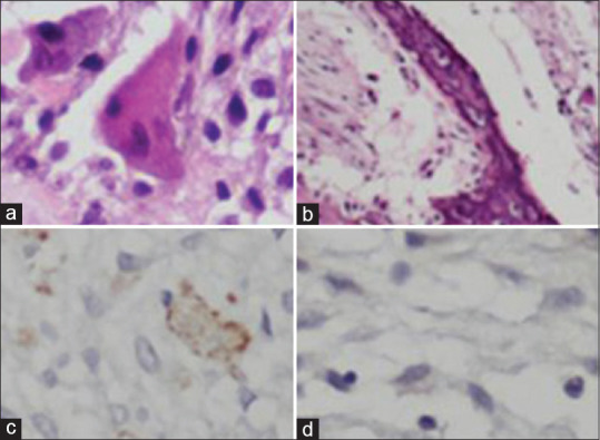
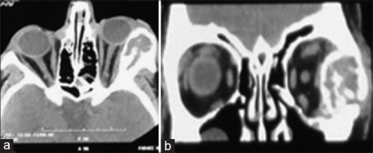
Multislice computed tomography scan (CT scan) of the orbit and paranasal sinuses showed a hyperdense, oval, extraconal mass with bone erosion. Magnetic resonance imaging of the orbit showed an inferior lateral isointense, oval, extraconal mass that had indented the globe. The patient underwent superior lateral orbitotomy, and the orbital mass was excised. Two months later, the patient developed proptosis, severe chemosis, and eyelid erythema in the same eye. CT scan showed an intraosseous mass in the lateral wall of the orbit that had pushed the globe anteromedially. Intraosseous tumor was resected, and the lateral orbital wall was drilled during the second surgery. GCT was diagnosed based on pathological survey.

CONCLUSION

Following the resection of the orbital GCT, the tumor behavior may change to an intraosseous lesion.

摘要

目的

报告1例眼眶骨巨细胞瘤(GCT),其表现从眶内肿块转变为骨内肿瘤。

方法

一名16岁男孩出现左上、下眼睑疼痛、肿胀、红斑、眼球突出和复视。眼科检查发现左眼结膜水肿、结膜充血、上抬受限、下转受限以及外展受限。

结果

眼眶和鼻窦的多层计算机断层扫描(CT扫描)显示一个高密度椭圆形眶锥外肿块伴有骨质侵蚀。眼眶磁共振成像显示一个位于下外侧等信号的椭圆形眶锥外肿块,该肿块使眼球受压。患者接受了眶上外侧切开术,切除了眼眶肿块。两个月后,患者同一只眼睛再次出现眼球突出、严重结膜水肿和眼睑红斑。CT扫描显示眼眶外侧壁有一个骨内肿块,将眼球向前内侧推移。第二次手术切除了骨内肿瘤,并对眼眶外侧壁进行了钻孔。根据病理检查诊断为骨巨细胞瘤。

结论

眼眶骨巨细胞瘤切除后,肿瘤表现可能转变为骨内病变。

https://cdn.ncbi.nlm.nih.gov/pmc/blobs/a7e2/7861103/c74d4f9fd547/JCO-32-414-g004.jpg
https://cdn.ncbi.nlm.nih.gov/pmc/blobs/a7e2/7861103/fefe0de923eb/JCO-32-414-g001.jpg
https://cdn.ncbi.nlm.nih.gov/pmc/blobs/a7e2/7861103/151646c0a0b4/JCO-32-414-g002.jpg
https://cdn.ncbi.nlm.nih.gov/pmc/blobs/a7e2/7861103/9848c292caec/JCO-32-414-g003.jpg
https://cdn.ncbi.nlm.nih.gov/pmc/blobs/a7e2/7861103/c74d4f9fd547/JCO-32-414-g004.jpg

相似文献

1
Giant Cell Tumor: Changing Behavior from Intraorbital to Intraosseous Mass.骨巨细胞瘤:从眶内肿物到骨内肿物的行为变化
J Curr Ophthalmol. 2020 Dec 12;32(4):414-416. doi: 10.4103/JOCO.JOCO_63_20. eCollection 2020 Oct-Dec.
2
Intraosseous meningioma of the sphenoid bone.蝶骨骨内脑膜瘤
Radiol Case Rep. 2015 Nov 6;5(1):357. doi: 10.2484/rcr.v5i1.357. eCollection 2010.
3
Ocular scrofuloderma with unilateral proptosis.伴有单侧眼球突出的眼皮肤结核。
J Dermatol. 2002 Apr;29(4):232-4. doi: 10.1111/j.1346-8138.2002.tb00255.x.
4
[Intraorbital schwannoma arising from the supraorbital nerve].[起源于眶上神经的眶内神经鞘瘤]
No Shinkei Geka. 2012 Feb;40(2):167-71.
5
Intraorbital Dermoid Cyst With Zygomaticofrontal Suture Erosion in an Adult.成人眶内皮样囊肿伴颧额缝侵蚀
J Craniofac Surg. 2019 Mar/Apr;30(2):514-515. doi: 10.1097/SCS.0000000000005005.
6
Orbital myopericytoma: an unusual tumor in an unusual location.眶内肌纤维母细胞瘤:不常见部位的不常见肿瘤。
Orbit. 2022 Aug;41(4):502-505. doi: 10.1080/01676830.2021.1890135. Epub 2021 Feb 26.
7
Giant Cell Tumor of Bone, Rare Inferior Orbit Location.骨巨细胞瘤,罕见的眶下位置
Plast Reconstr Surg Glob Open. 2024 Feb 1;12(2):e5569. doi: 10.1097/GOX.0000000000005569. eCollection 2024 Feb.
8
Atypical sino-orbital inflammatory myofibroblastic tumor with bone and cerebral invasion extending to the orbit.伴有骨质及脑侵犯并累及眼眶的非典型鼻窦-眼眶炎性肌纤维母细胞瘤。
Eur J Ophthalmol. 2014 Jul-Aug;24(4):608-10. doi: 10.5301/ejo.5000401. Epub 2013 Dec 9.
9
Intraorbital haematoma during a commercial flight: a case report.商业航班中发生眶内血肿:病例报告。
BMC Ophthalmol. 2015 May 7;15:47. doi: 10.1186/s12886-015-0034-y.
10
Benign chondroid syringoma of the orbit: a rare cause of exophtalmos.眶部良性软骨样汗腺瘤:眼球突出的罕见原因。
Head Face Med. 2012 Mar 8;8:8. doi: 10.1186/1746-160X-8-8.

本文引用的文献

1
Giant cell tumour of frontal bone in a patient with bilateral retinoblastoma.
Arch Soc Esp Oftalmol (Engl Ed). 2019 Apr;94(4):200-203. doi: 10.1016/j.oftal.2018.10.003. Epub 2018 Nov 16.
2
Denosumab in Patients with Giant Cell Tumor and Its Recurrence: A Systematic Review.地诺单抗治疗骨巨细胞瘤及其复发患者的系统评价
Arch Bone Jt Surg. 2018 Jul;6(4):260-268.
3
Left orbital roof giant cell tumor of bone: A case report.左眼眶顶骨巨细胞瘤:一例报告。
Surg Neurol Int. 2018 Jun 26;9:127. doi: 10.4103/sni.sni_467_17. eCollection 2018.
4
Giant Cell Tumor of the Lateral Skull Base: Diagnostic and Management Options.外侧颅底巨细胞瘤:诊断与治疗选择
J Neurol Surg Rep. 2018 Apr;79(2):e41-e54. doi: 10.1055/s-0038-1645885. Epub 2018 May 25.
5
Denosumab treatment for progressive skull base giant cell tumor of bone in a 14 year old female - a case report and literature review.地诺单抗治疗一名14岁女性进展性颅底骨巨细胞瘤——病例报告及文献综述
Ital J Pediatr. 2017 Mar 29;43(1):32. doi: 10.1186/s13052-017-0353-0.
6
Giant Cell Tumor of the Frontal Bone Presenting as an Orbital Mass.表现为眼眶肿物的额骨巨细胞瘤
Ophthalmol Ther. 2017 Jun;6(1):215-220. doi: 10.1007/s40123-017-0081-y. Epub 2017 Feb 16.
7
Treatment of a skull-base giant cell tumor with endoscopic endonasal resection and denosumab: case report.内镜经鼻颅底巨大细胞瘤切除术联合地舒单抗治疗:病例报告。
J Neurosurg. 2017 Feb;126(2):431-434. doi: 10.3171/2016.3.JNS152802. Epub 2016 May 13.
8
Giant Cell Tumor of Bone - An Overview.骨巨细胞瘤——概述
Arch Bone Jt Surg. 2016 Jan;4(1):2-9.
9
Giant Cell Tumor of the Skull: Review of the Literature.颅骨巨细胞瘤:文献综述
J Neurol Surg A Cent Eur Neurosurg. 2016 May;77(3):239-46. doi: 10.1055/s-0035-1554808. Epub 2015 Jun 19.
10
RANKL, denosumab, and giant cell tumor of bone.RANKL、地舒单抗和骨巨细胞瘤。
Curr Opin Oncol. 2012 Jul;24(4):397-403. doi: 10.1097/CCO.0b013e328354c129.