Syracuse University, Department of Biology, 107 College Place, Syracuse, NY 13244, USA.
STAR Protoc. 2021 Jan 22;2(1):100293. doi: 10.1016/j.xpro.2020.100293. eCollection 2021 Mar 19.
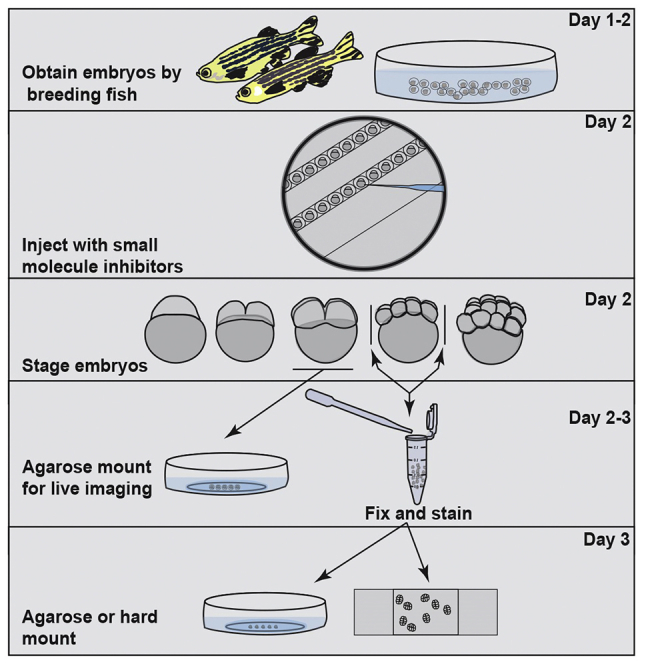
During the earliest division stages, zebrafish embryos have large cells that divide rapidly and synchronously to create a cellular layer on top of the yolk. Here, we describe a protocol for monitoring spindle dynamics during these early embryonic divisions. We outline techniques for injecting zebrafish embryos with small-molecule inhibitors toward polo-like kinases, preparing and mounting embryos for three-dimensional imaging using confocal microscopy. These techniques are used to understand how the early zebrafish embryo's centrosome constructs the mitotic spindle. For complete details on the use and execution of this protocol, please refer to Rathbun et al. (2020).
在最早的分裂阶段,斑马鱼胚胎具有快速分裂且同步的大细胞,从而在蛋黄上方形成一层细胞。在这里,我们描述了一种监测这些早期胚胎分裂过程中纺锤体动态的方案。我们概述了向斑马鱼胚胎注射小分子抑制剂以抑制 Polo 样激酶的技术,以及准备和安装胚胎以进行共聚焦显微镜的三维成像的技术。这些技术用于了解早期斑马鱼胚胎的中心体如何构建有丝分裂纺锤体。有关此方案的使用和执行的完整详细信息,请参阅 Rathbun 等人。(2020 年)。