Department of Pharmacology, University of North Carolina at Chapel Hill, Chapel Hill, NC 27599, USA.
Neuroscience Center, University of North Carolina at Chapel Hill, Chapel Hill, NC 27599, USA.
STAR Protoc. 2021 Feb 1;2(1):100306. doi: 10.1016/j.xpro.2021.100306. eCollection 2021 Mar 19.
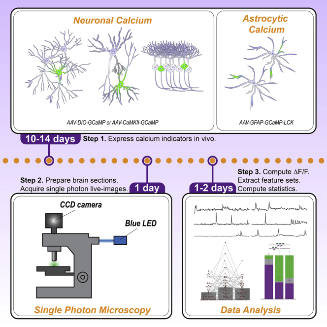
Confocal, multiphoton, or other advanced microscopy techniques produce high-quality datasets of calcium activity in live tissue. However, researchers without access to such expensive equipment can still produce meaningful observations from single-photon datasets. Here, we describe a protocol to extract meaningful features of both somatic neuronal and membranous astrocytic calcium dynamics obtained from charge-coupled device (CCD)-based camera setups, typical of electrophysiology rigs and highly relevant for investigating neuronal and astrocytic involvement in brain circuitry. For complete details on the use and execution of this protocol, please refer to Asrican et al. (2020).
共聚焦、多光子或其他先进的显微镜技术可生成活组织中钙活性的高质量数据集。然而,没有使用这种昂贵设备的研究人员仍然可以从单光子数据集获得有意义的观察结果。在这里,我们描述了一种从基于电荷耦合器件 (CCD) 的相机设置中提取体细胞神经元和膜性星形胶质细胞钙动力学有意义特征的方案,该方案典型地用于电生理学装置,对于研究神经元和星形胶质细胞在大脑回路中的参与非常相关。有关此协议的使用和执行的完整详细信息,请参阅 Asrican 等人。(2020 年)。