Centre de Résonance Magnétique des Systèmes Biologiques, UMR 5536, CNRS/Univ. Bordeaux, 146 rue Léo Saignat, 33076, Bordeaux, France.
Department of General, Organic and Biomedical Chemistry, NMR and Molecular Imaging Laboratory, University of Mons, 19 avenue Maistriau, 7000, Mons, Belgium.
Sci Rep. 2021 Feb 8;11(1):3286. doi: 10.1038/s41598-021-82095-6.
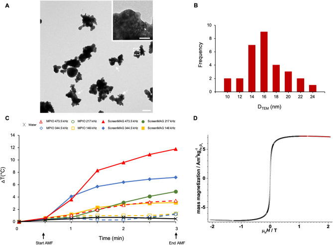
Iron oxide particles (IOP) are commonly used for Cellular Magnetic Resonance Imaging (MRI) and in combination with several treatments, like Magnetic Fluid Hyperthermia (MFH), due to the rise in temperature they provoke under an Alternating Magnetic Field (AMF). Micrometric IOP have a high sensitivity of detection. Nevertheless, little is known about their internalization processes or their potential heat power. Two micrometric commercial IOP (from Bangs Laboratories and Chemicell) were characterized by Transmission Electron Microscopy (TEM) and their endocytic pathways into glioma cells were analyzed. Their Specific Absorption Rate (SAR) and cytotoxicity were evaluated using a commercial AMF inductor. T2-weighted imaging was used to monitor tumor growth in vivo after MFH treatment in mice. The two micron-sized IOP had similar structures and r relaxivities (100 mM s) but involved different endocytic pathways. Only ScreenMAG particles generated a significant rise in temperature following AMF (SAR = 113 W g Fe). After 1 h of AMF exposure, 60% of ScreenMAG-labeled cells died. Translated to a glioma model, 89% of mice responded to the treatment with smaller tumor volume 42 days post-implantation. Micrometric particles were investigated from their characterization to their intracellular internalization pathways and applied in one in vivo cancer treatment, i.e. MFH.
氧化铁粒子(IOP)常用于细胞磁共振成像(MRI),并与多种治疗方法结合使用,如磁流体热疗(MFH),因为它们在交变磁场(AMF)下会引起温度升高。微米级的 IOP 具有很高的检测灵敏度。然而,对于它们的内化过程或潜在的发热能力,人们知之甚少。两种商业微米级 IOP(来自 Bangs 实验室和 ChemiCell)通过透射电子显微镜(TEM)进行了表征,并分析了它们进入神经胶质瘤细胞的内吞途径。使用商用 AMF 感应器评估了它们的比吸收率(SAR)和细胞毒性。在 T2 加权成像中,在 MFH 治疗后监测了体内肿瘤的生长。两种微米级 IOP 具有相似的结构和 r2弛豫率(100mM s),但涉及不同的内吞途径。只有 ScreenMAG 颗粒在 AMF 后会产生显著的温升(SAR=113W g Fe)。在 AMF 暴露 1 小时后,60%的 ScreenMAG 标记细胞死亡。在神经胶质瘤模型中,89%的小鼠对治疗有反应,植入后 42 天肿瘤体积更小。从特性到细胞内内化途径,对微米级颗粒进行了研究,并应用于一种体内癌症治疗,即 MFH。