Department of Neurosurgery, West Virginia University, 1 Medical Center Drive, Suite 4300, Morgantown, WV, 26506-9183, USA.
Department of Radiation Oncology, West Virginia University, Morgantown, WV, USA.
J Neurooncol. 2021 Apr;152(2):245-255. doi: 10.1007/s11060-021-03707-9. Epub 2021 Feb 10.
Radiotherapy-induced tumor death remains critical in the successful first-line management of glioblastoma, whereas resistance to radiation serves as a major factor in disease progression. Mesenchymal shift has been identified as a driver in GBM recurrence, with gene expression associated with enhanced repair of macromolecular damage caused by radiation.
Using distinct mesenchymal subtype GBM cells lines, radiation response was assessed by clonogenic assay and orthotopic mouse tumor model. RNA-sequencing was performed in the setting of increasing radiation dosing while real-time assessment of ROS generation was achieved by the measurement of hydroxyl spin trap adducts via electron paramagnetic resonance.
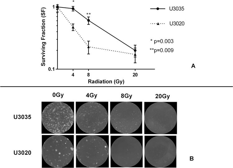
Radiation-induced cell death determined by clonogenic assay was significantly different at low dose (4-8 Gy) between the resistant U3035 cells and the sensitive U3020 cells. Similar trends were present in the in vivo NSG mouse model following radiation dosing on post-implantation day 7-10, with the rate of reduction in tumor bioluminescence reversing between the U3020 and U3035 cells after the third dose of radiation. Changes in gene expression following radiation determined by RNA-sequencing indicate both U3035 and U3020 cells demonstrate a shift toward more mesenchymal profiles, with concurrent shift away from pro-neural subtype gene expression in the U3020 cells that appeared to develop resistance to radiation in vivo. Persistence of ROS generated following radiation was greater in U3020 cells shown to be more sensitive to radiation.
Despite the same molecular classification, distinct GBM cell lines can demonstrate differential response to radiation and potential for mesenchymal shift associated with radiation resistance.
放射治疗诱导的肿瘤死亡仍然是胶质母细胞瘤成功一线治疗的关键,而对辐射的抵抗是疾病进展的主要因素。间充质转移已被确定为 GBM 复发的驱动因素,其基因表达与增强辐射引起的大分子损伤修复有关。
使用不同的间充质亚型 GBM 细胞系,通过集落形成测定和原位小鼠肿瘤模型评估放射反应。在增加放射剂量的情况下进行 RNA 测序,同时通过电子顺磁共振测量羟基自旋捕获加合物来实时评估 ROS 生成。
集落形成测定确定的低剂量(4-8Gy)下,耐药 U3035 细胞与敏感 U3020 细胞之间的放射诱导细胞死亡有显著差异。在放射后第 7-10 天植入后的 NSG 小鼠模型中也存在类似的趋势,在第 3 次放射后,U3020 和 U3035 细胞之间的肿瘤生物发光减少率发生逆转。RNA 测序确定的放射后基因表达变化表明,U3035 和 U3020 细胞均表现出向更间充质表型的转变,同时 U3020 细胞中向神经前体亚型基因表达的转变也发生了转变,这似乎使其在体内对放射产生了耐药性。在对放射更敏感的 U3020 细胞中,放射后生成的 ROS 持续时间更长。
尽管具有相同的分子分类,但不同的 GBM 细胞系对放射的反应和潜在的与放射抵抗相关的间充质转移能力可能不同。