• 文献检索
  • 文档翻译
  • 深度研究
  • 学术资讯
  • Suppr Zotero 插件Zotero 插件
  • 邀请有礼
  • 套餐&价格
  • 历史记录
应用&插件
Suppr Zotero 插件Zotero 插件浏览器插件Mac 客户端Windows 客户端微信小程序
定价
高级版会员购买积分包购买API积分包
服务
文献检索文档翻译深度研究API 文档MCP 服务
关于我们
关于 Suppr公司介绍联系我们用户协议隐私条款
关注我们

Suppr 超能文献

核心技术专利:CN118964589B侵权必究
粤ICP备2023148730 号-1Suppr @ 2026

文献检索

告别复杂PubMed语法,用中文像聊天一样搜索,搜遍4000万医学文献。AI智能推荐,让科研检索更轻松。

立即免费搜索

文件翻译

保留排版,准确专业,支持PDF/Word/PPT等文件格式,支持 12+语言互译。

免费翻译文档

深度研究

AI帮你快速写综述,25分钟生成高质量综述,智能提取关键信息,辅助科研写作。

立即免费体验

食管局部晚期腺癌中肿瘤浸润性T淋巴细胞的分布及避免免疫细胞攻击的可能肿瘤逃逸机制

Distribution of tumor-infiltrating-T-lymphocytes and possible tumor-escape mechanisms avoiding immune cell attack in locally advanced adenocarcinomas of the esophagus.

作者信息

Schoemmel M, Loeser H, Kraemer M, Wagener-Ryczek S, Hillmer A, Bruns C, Thelen M, Schröder W, Zander T, Lechner A, Buettner R, Schlösser H, Gebauer F, Quaas A

机构信息

Institute of Pathology, University-Hospital of Cologne, University of Cologne, Kerpener Strasse 62, 50937, Cologne, Germany.

Department of General, Visceral and Cancer Surgery, University of Cologne, Cologne, Germany.

出版信息

Clin Transl Oncol. 2021 Aug;23(8):1601-1610. doi: 10.1007/s12094-021-02556-2. Epub 2021 Feb 10.

DOI:10.1007/s12094-021-02556-2
PMID:33566304
原文链接:https://pmc.ncbi.nlm.nih.gov/articles/PMC8238763/
Abstract

INTRODUCTION

The inflammatory microenvironment has emerged as one of the focuses of cancer research. Little is known about the immune environment in esophageal adenocarcinoma (EAC) and possible tumor-escape mechanisms to avoid immune cell attack.

PATIENTS AND METHODS

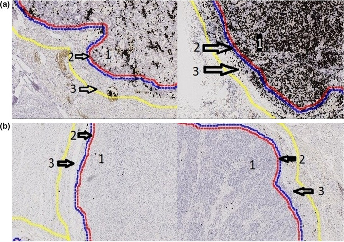
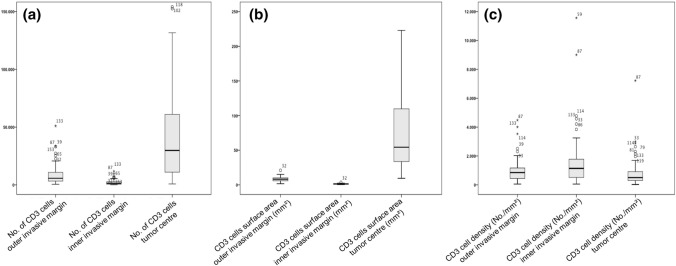
We measured T cell inflammation (CD3, CD8) in the microenvironment using a standardized software-based evaluation algorithm considering different predefined tumor areas as well as expression of MHC class 1 and PD-L1 on 75 analyzable primarily resected and locally advanced (≥ pT2) EACs. We correlated these findings statistically with clinical data.

RESULTS

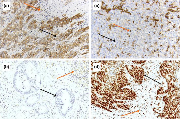
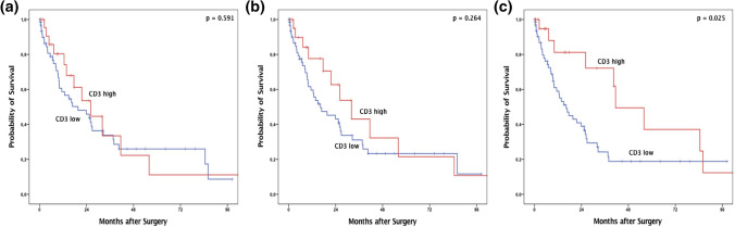
Patients with high amounts of T cell infiltration in their tumor center showed a significant survival benefit of 41.4 months compared to 16.3 months in T cell poor tumors (p = 0.025), although CD3 fails to serve as an independent prognostic marker in multivariate analysis. For the invasion zone, a correlation between number of T-cells and overall survival was not detectable. Loss of MHC1 protein expression on tumor cells was seen in 32% and PD-L1 expression using the combined positive score (CPS) in 21.2%. Most likely due to small numbers of cases, both markers are not prognostically relevant, even though PD-L1 expression correlates with advanced tumor stages.

DISCUSSION

Our analyses reveal an outstanding, though not statistically independent, prognostic relevance of T-cell-rich inflammation in our group of EACs, in particular driven by the tumor center. For the first time, we describe that the inner part of the invasion zone in EACs shows significantly fewer T-cells than other tumor segments and is prognostically irrelevant. We also demonstrate that the loss of antigen presenting ability via MHC1 downregulation by the carcinoma cells is a common escape mechanism in EACs. Future work will need to show whether tumors with MHC class 1 loss respond less well to immunotherapy.

摘要

引言

炎症微环境已成为癌症研究的重点之一。关于食管腺癌(EAC)中的免疫环境以及避免免疫细胞攻击的可能肿瘤逃逸机制,我们所知甚少。

患者与方法

我们使用基于软件的标准化评估算法,在微环境中测量T细胞炎症(CD3、CD8),该算法考虑了不同的预定义肿瘤区域,以及75例可分析的原发性切除和局部晚期(≥pT2)EAC中MHC 1类和PD-L1的表达。我们将这些发现与临床数据进行了统计学关联。

结果

肿瘤中心T细胞浸润量高的患者显示出显著的生存获益,中位生存期为41.4个月,而T细胞浸润少的肿瘤患者为16.3个月(p = 0.025),尽管在多变量分析中CD3不能作为独立的预后标志物。对于浸润区,未检测到T细胞数量与总生存期之间的相关性。32%的肿瘤细胞出现MHC1蛋白表达缺失,使用联合阳性评分(CPS)时PD-L1表达为21.2%。很可能由于病例数较少,这两个标志物均无预后相关性,尽管PD-L1表达与肿瘤晚期相关。

讨论

我们的分析揭示了在我们的EAC队列中,富含T细胞的炎症具有显著的预后相关性,尽管在统计学上不独立,尤其是由肿瘤中心驱动。我们首次描述,EAC浸润区内部的T细胞明显少于其他肿瘤节段,且无预后相关性。我们还证明,癌细胞通过下调MHC1导致抗原呈递能力丧失是EAC中常见的逃逸机制。未来的研究需要表明,MHC 1缺失的肿瘤对免疫治疗的反应是否较差。

https://cdn.ncbi.nlm.nih.gov/pmc/blobs/43f4/8238763/506319f7e52e/12094_2021_2556_Fig4_HTML.jpg
https://cdn.ncbi.nlm.nih.gov/pmc/blobs/43f4/8238763/6fc00b1fb0dc/12094_2021_2556_Fig1_HTML.jpg
https://cdn.ncbi.nlm.nih.gov/pmc/blobs/43f4/8238763/dc4ad4dc17de/12094_2021_2556_Fig2_HTML.jpg
https://cdn.ncbi.nlm.nih.gov/pmc/blobs/43f4/8238763/28260a4e4722/12094_2021_2556_Fig3_HTML.jpg
https://cdn.ncbi.nlm.nih.gov/pmc/blobs/43f4/8238763/506319f7e52e/12094_2021_2556_Fig4_HTML.jpg

相似文献

1
Distribution of tumor-infiltrating-T-lymphocytes and possible tumor-escape mechanisms avoiding immune cell attack in locally advanced adenocarcinomas of the esophagus.食管局部晚期腺癌中肿瘤浸润性T淋巴细胞的分布及避免免疫细胞攻击的可能肿瘤逃逸机制
Clin Transl Oncol. 2021 Aug;23(8):1601-1610. doi: 10.1007/s12094-021-02556-2. Epub 2021 Feb 10.
2
PD-L1 expression in carcinoma of the esophagogastric junction is positively correlated with T-cell infiltration and overall survival.食管胃结合部癌中 PD-L1 的表达与 T 细胞浸润和总生存呈正相关。
Pathol Res Pract. 2019 Jun;215(6):152402. doi: 10.1016/j.prp.2019.03.030. Epub 2019 Apr 20.
3
PD-L1 Expression in Endocervical Adenocarcinoma: Correlation With Patterns of Tumor Invasion, CD8+ Tumor-infiltrating Lymphocytes, and Clinical Outcomes.PD-L1 表达在内膜腺癌中的表现:与肿瘤浸润模式、CD8+肿瘤浸润淋巴细胞和临床结局的相关性。
Am J Surg Pathol. 2021 Jun 1;45(6):742-752. doi: 10.1097/PAS.0000000000001633.
4
Tumor-Infiltrating Immune Cells and PD-L1 as Prognostic Biomarkers in Primary Esophageal Small Cell Carcinoma.肿瘤浸润免疫细胞和 PD-L1 作为原发性食管小细胞癌的预后生物标志物。
J Immunol Res. 2020 Dec 29;2020:8884683. doi: 10.1155/2020/8884683. eCollection 2020.
5
Increased intraepithelial CD3+ T-lymphocytes and high PD-L1 expression on tumor cells are associated with a favorable prognosis in esophageal squamous cell carcinoma and allow prognostic immunogenic subgrouping.上皮内CD3+ T淋巴细胞增多以及肿瘤细胞上程序性死亡受体配体1(PD-L1)高表达与食管鳞状细胞癌的良好预后相关,并可进行预后免疫原性亚组划分。
Oncotarget. 2017 Jul 18;8(29):46756-46768. doi: 10.18632/oncotarget.18606.
6
Prognostic perspectives of PD-L1 combined with tumor-infiltrating lymphocytes, Epstein-Barr virus, and microsatellite instability in gastric carcinomas.胃腺癌中 PD-L1 联合肿瘤浸润淋巴细胞、EB 病毒和微卫星不稳定性的预后预测。
Diagn Pathol. 2020 Jun 4;15(1):69. doi: 10.1186/s13000-020-00979-z.
7
Programmed Death Ligand 1: A Step Toward Immunoscore for Esophageal Cancer.程序性死亡配体 1:迈向食管癌免疫评分的一步。
Ann Thorac Surg. 2018 Oct;106(4):1002-1007. doi: 10.1016/j.athoracsur.2018.05.002. Epub 2018 May 30.
8
Differential expression of PD-L1 and IDO1 in association with the immune microenvironment in resected lung adenocarcinomas.肺腺癌切除标本中 PD-L1 和 IDO1 的表达差异与免疫微环境的关系。
Mod Pathol. 2019 Apr;32(4):511-523. doi: 10.1038/s41379-018-0160-1. Epub 2018 Oct 26.
9
Immune microenvironment and evasion mechanisms in adenoid cystic carcinomas of salivary glands.涎腺腺样囊性癌的免疫微环境与逃逸机制。
Oral Oncol. 2019 Jan;88:95-101. doi: 10.1016/j.oraloncology.2018.11.028. Epub 2018 Nov 23.
10
The prognostic impact of programmed cell death ligand 1 and human leukocyte antigen class I in pancreatic cancer.程序性细胞死亡配体1和人类白细胞抗原I类在胰腺癌中的预后影响。
Cancer Med. 2017 Jul;6(7):1614-1626. doi: 10.1002/cam4.1087. Epub 2017 Jun 10.

引用本文的文献

1
Tumor-Infiltrating Lymphocytes Demonstrate Distinct Behavior in the Tumoral and Peritumoral Microenvironment After Neoadjuvant Chemotherapy in Patients with Resected Adenocarcinoma of the Gastric or Gastroesophageal Junction: Results from a Single Center.肿瘤浸润淋巴细胞在接受新辅助化疗后的胃或胃食管交界腺癌切除患者的肿瘤及瘤周微环境中表现出不同行为:单中心研究结果
Cancers (Basel). 2025 Jun 13;17(12):1971. doi: 10.3390/cancers17121971.
2
STAT5 Activation Enhances Adoptive Therapy Combined with Peptide Vaccination by Preventing PD-1 Inhibition.信号转导和转录激活因子5(STAT5)的激活通过防止程序性死亡受体1(PD-1)抑制作用增强了过继性疗法与肽疫苗接种相结合的效果。
Mol Cancer Ther. 2025 Mar 4;24(3):419-430. doi: 10.1158/1535-7163.MCT-24-0505.
3

本文引用的文献

1
Immune microenvironment in Barrett's esophagus adjacent to esophageal adenocarcinoma: possible influence of adjacent mucosa on cancer development and progression.巴雷特食管毗邻食管腺癌的免疫微环境:毗邻黏膜对癌症发生和发展的可能影响。
Virchows Arch. 2020 Dec;477(6):825-834. doi: 10.1007/s00428-020-02854-0. Epub 2020 Jun 12.
2
Hybrid Minimally Invasive Esophagectomy for Esophageal Cancer.杂交微创食管癌切除术。
N Engl J Med. 2019 Jan 10;380(2):152-162. doi: 10.1056/NEJMoa1805101.
3
Genomics of response to immune checkpoint therapies for cancer: implications for precision medicine.
Tumor-Infiltrating Lymphocytes in Resected Esophageal and Gastric Adenocarcinomas Do Not Correlate with Tumor Regression Score After Neoadjuvant Chemotherapy: Results of a Case-Series Study.
切除的食管腺癌和胃腺癌中的肿瘤浸润淋巴细胞与新辅助化疗后的肿瘤退缩评分不相关:一项病例系列研究的结果
Cancers (Basel). 2024 Nov 1;16(21):3694. doi: 10.3390/cancers16213694.
4
Analyzing risk factors and constructing a predictive model for superficial esophageal carcinoma with submucosal infiltration exceeding 200 micrometers.分析黏膜下浸润超过 200 微米的表浅食管癌的危险因素并构建预测模型。
BMC Gastroenterol. 2024 Oct 6;24(1):350. doi: 10.1186/s12876-024-03442-1.
5
Cell cycle associated protein 1 associates with immune infiltration and ferroptosis in gastrointestinal cancer.细胞周期相关蛋白1与胃肠道癌中的免疫浸润和铁死亡相关。
Heliyon. 2024 Mar 27;10(7):e28794. doi: 10.1016/j.heliyon.2024.e28794. eCollection 2024 Apr 15.
6
Tumour microenvironment influences response to treatment in oesophageal adenocarcinoma.肿瘤微环境影响食管腺癌的治疗反应。
Front Immunol. 2023 Dec 13;14:1330635. doi: 10.3389/fimmu.2023.1330635. eCollection 2023.
7
Establish immune-related gene prognostic index for esophageal cancer.建立食管癌免疫相关基因预后指数。
Front Genet. 2022 Aug 9;13:956915. doi: 10.3389/fgene.2022.956915. eCollection 2022.
8
Fructose-1,6-bisphosphatase 1 (FBP1) is an independent biomarker associated with a favorable prognosis in esophageal adenocarcinoma.果糖-1,6-二磷酸酶 1(FBP1)是食管腺癌预后良好的独立生物标志物。
J Cancer Res Clin Oncol. 2022 Sep;148(9):2287-2293. doi: 10.1007/s00432-022-04025-x. Epub 2022 Apr 27.
9
C5b-9 Membrane Attack Complex Formation and Extracellular Vesicle Shedding in Barrett's Esophagus and Esophageal Adenocarcinoma.巴雷特食管和食管腺癌中C5b-9膜攻击复合物的形成及细胞外囊泡脱落
Front Immunol. 2022 Mar 8;13:842023. doi: 10.3389/fimmu.2022.842023. eCollection 2022.
癌症免疫检查点治疗反应的基因组学:对精准医学的启示。
Genome Med. 2018 Nov 29;10(1):93. doi: 10.1186/s13073-018-0605-7.
4
A systematic review and meta-analysis of prognostic biomarkers in resectable esophageal adenocarcinomas.可切除食管腺癌中预后生物标志物的系统评价和荟萃分析。
Sci Rep. 2018 Sep 5;8(1):13281. doi: 10.1038/s41598-018-31548-6.
5
Clinical Utility of the Combined Positive Score for Programmed Death Ligand-1 Expression and the Approval of Pembrolizumab for Treatment of Gastric Cancer.程序性死亡配体-1 表达联合阳性评分在帕博利珠单抗治疗胃癌中的临床应用价值。
Arch Pathol Lab Med. 2019 Mar;143(3):330-337. doi: 10.5858/arpa.2018-0043-OA. Epub 2018 Jul 20.
6
MHC proteins confer differential sensitivity to CTLA-4 and PD-1 blockade in untreated metastatic melanoma.MHC 蛋白赋予未经治疗的转移性黑色素瘤对 CTLA-4 和 PD-1 阻断的不同敏感性。
Sci Transl Med. 2018 Jul 18;10(450). doi: 10.1126/scitranslmed.aar3342.
7
PD-L1 expression is an independent predictor of favorable outcome in patients with localized esophageal adenocarcinoma.程序性死亡受体配体1(PD-L1)表达是局限性食管腺癌患者预后良好的独立预测指标。
Oncoimmunology. 2018 Mar 13;7(6):e1435226. doi: 10.1080/2162402X.2018.1435226. eCollection 2018.
8
Comparative Molecular Analyses of Esophageal Squamous Cell Carcinoma, Esophageal Adenocarcinoma, and Gastric Adenocarcinoma.食管鳞癌、食管腺癌和胃腺癌的比较分子分析。
Oncologist. 2018 Nov;23(11):1319-1327. doi: 10.1634/theoncologist.2018-0143. Epub 2018 Jun 4.
9
Programmed Death Ligand 1: A Step Toward Immunoscore for Esophageal Cancer.程序性死亡配体 1:迈向食管癌免疫评分的一步。
Ann Thorac Surg. 2018 Oct;106(4):1002-1007. doi: 10.1016/j.athoracsur.2018.05.002. Epub 2018 May 30.
10
Epstein-Barr virus and mismatch repair deficiency status differ between oesophageal and gastric cancer: A large multi-centre study.爱泼斯坦-巴尔病毒和错配修复缺陷状态在食管癌和胃癌之间存在差异:一项大型多中心研究。
Eur J Cancer. 2018 May;94:104-114. doi: 10.1016/j.ejca.2018.02.014. Epub 2018 Mar 20.