Zong Hailing, Hazelbaker Mark, Moe Christina, Ems-McClung Stephanie C, Hu Ke, Walczak Claire E
Department of Biology, Indiana University, Bloomington, IN 47405.
Medical Sciences, Indiana University School of Medicine-Bloomington, Bloomington, IN 47405.
Mol Biol Cell. 2021 Apr 1;32(7):590-604. doi: 10.1091/mbc.E20-05-0301. Epub 2021 Feb 10.
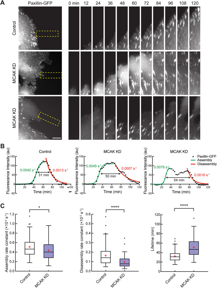
The asymmetric distribution of microtubule (MT) dynamics in migrating cells is important for cell polarization, yet the underlying regulatory mechanisms remain underexplored. Here, we addressed this question by studying the role of the MT depolymerase, MCAK (mitotic centromere-associated kinesin), in the highly persistent migration of RPE-1 cells. MCAK knockdown leads to slowed migration and poor directional movement. Fixed and live cell imaging revealed that MCAK knockdown results in excessive membrane ruffling as well as defects in cell polarization and the maintenance of a major protrusive front. Additionally, loss of MCAK increases the lifetime of focal adhesions by decreasing their disassembly rate. These functions correlate with a spatial distribution of MCAK activity, wherein activity is higher in the trailing edge of cells compared with the leading edge. Overexpression of Rac1 has a dominant effect over MCAK activity, placing it downstream of or in a parallel pathway to MCAK function in migration. Together, our data support a model in which the polarized distribution of MCAK activity and subsequent differential regulation of MT dynamics contribute to cell polarity, centrosome positioning, and focal adhesion dynamics, which all help facilitate robust directional migration.
微管(MT)动力学在迁移细胞中的不对称分布对细胞极化很重要,但其潜在的调控机制仍未得到充分研究。在这里,我们通过研究MT解聚酶MCAK(有丝分裂着丝粒相关驱动蛋白)在RPE - 1细胞高度持续迁移中的作用来解决这个问题。MCAK基因敲低导致迁移减慢和定向运动能力变差。固定和活细胞成像显示,MCAK基因敲低会导致过度的膜皱褶以及细胞极化缺陷和主要突出前沿的维持障碍。此外,MCAK的缺失通过降低粘着斑的解体速率来增加其寿命。这些功能与MCAK活性的空间分布相关,其中细胞后缘的活性高于前缘。Rac1的过表达对MCAK活性具有主导作用,使其在迁移过程中处于MCAK功能的下游或与之平行的途径中。总之,我们的数据支持一个模型,其中MCAK活性的极化分布以及随后对MT动力学的差异调节有助于细胞极性、中心体定位和粘着斑动力学,所有这些都有助于促进强劲的定向迁移。