Müller Manuel, Gorek Lena, Kamm Natalia, Jacob Ralf
Department of Cell Biology and Cell Pathology, Philipps-Universität Marburg, Marburg, Germany.
DFG Research Training Group, Membrane Plasticity in Tissue Development and Remodelling, GRK 2213, Philipps-Universität Marburg, Marburg, Germany.
Front Cell Dev Biol. 2022 Jul 12;10:901999. doi: 10.3389/fcell.2022.901999. eCollection 2022.
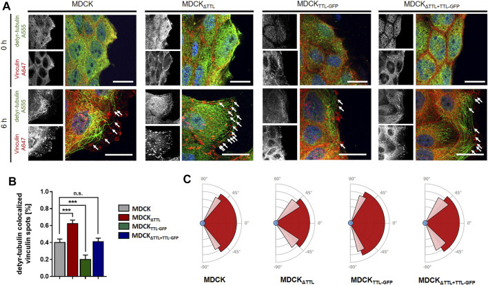
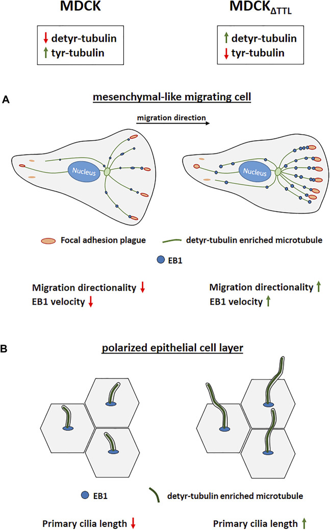
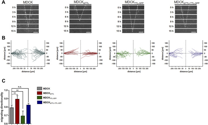
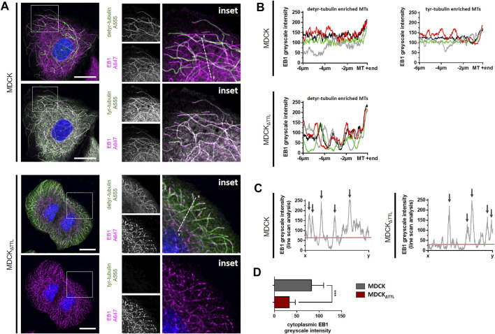
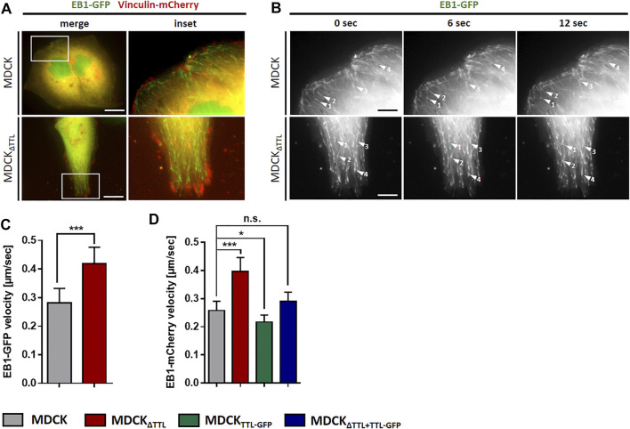
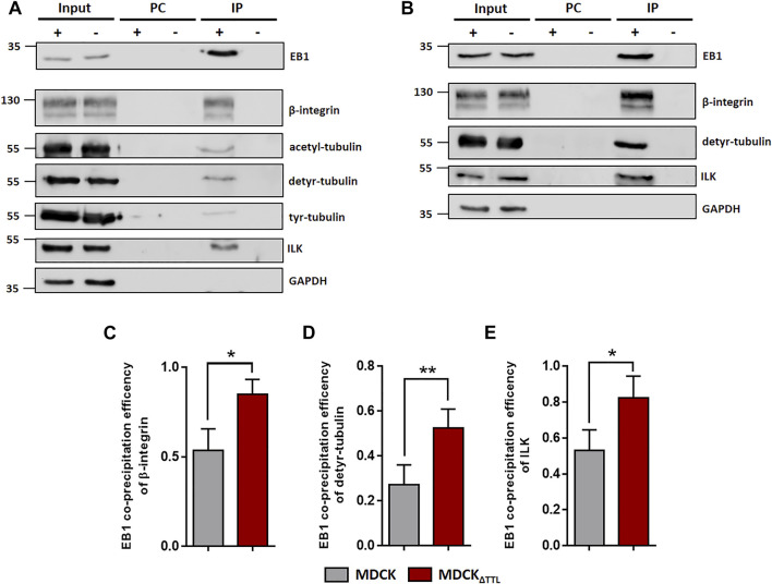
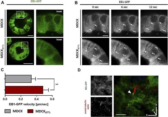
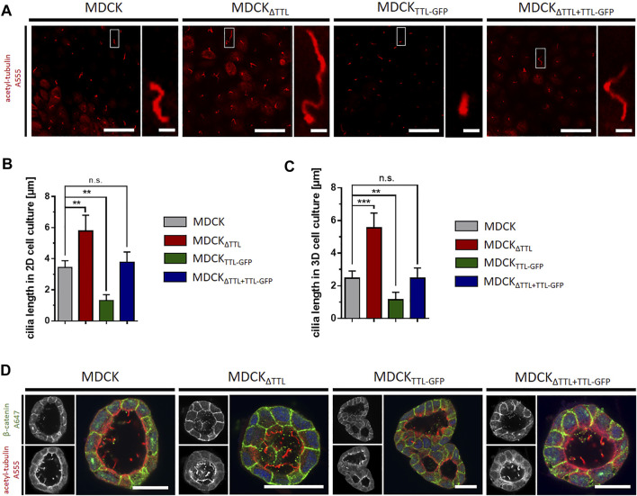
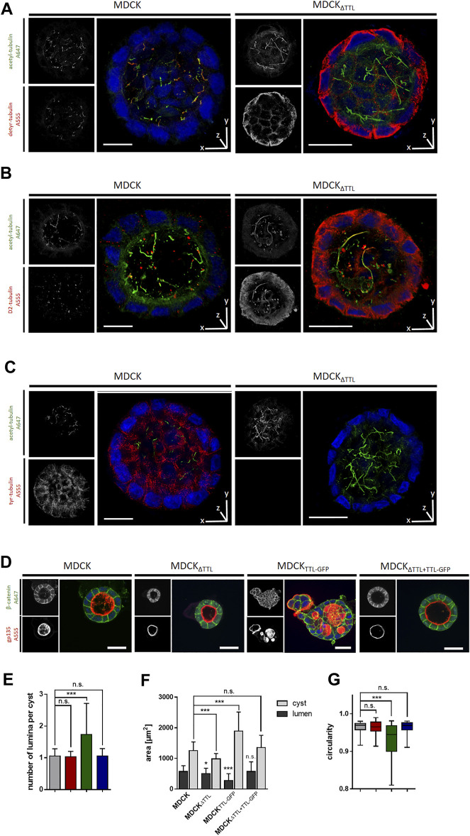
Conjunction of epithelial cells into monolayer sheets implies the ability to migrate and to undergo apicobasal polarization. Both processes comprise reorganization of cytoskeletal elements and rearrangements of structural protein interactions. We modulated expression of tubulin tyrosin ligase (TTL), the enzyme that adds tyrosine to the carboxy terminus of detyrosinated α-tubulin, to study the role of tubulin detyrosination/-tyrosination in the orientation of cell motility and in epithelial morphogenesis. Oriented cell migration and the organization of focal adhesions significantly lose directionality with diminishing amounts of microtubules enriched in detyrosinated tubulin. On the other hand, increasing quantities of detyrosinated tubulin results in faster plus end elongation of microtubules in migrating and in polarized epithelial cells. These plus ends are decorated by the plus end binding protein 1 (EB1), which mediates interaction between microtubules enriched in detyrosinated tubulin and the integrin-ILK complex at focal adhesions. EB1 accumulates at the apical cell pole at the base of the primary cilium following apicobasal polarization. Polarized cells almost devoid of detyrosinated tubulin form stunted primary cilia and multiluminal cysts in 3D-matrices. We conclude that the balance between detyrosinated and tyrosinated tubulin alters microtubule dynamics, affects the orientation of focal adhesions and determines the organization of primary cilia on epithelial cells.
上皮细胞连接形成单层片层意味着细胞具有迁移能力并能进行顶-基极化。这两个过程都包括细胞骨架成分的重组以及结构蛋白相互作用的重排。我们调节了微管蛋白酪氨酸连接酶(TTL)的表达,该酶可将酪氨酸添加到去酪氨酸化的α-微管蛋白的羧基末端,以研究微管蛋白去酪氨酸化/酪氨酸化在细胞运动方向和上皮形态发生中的作用。随着富含去酪氨酸化微管蛋白的微管数量减少,定向细胞迁移和粘着斑的组织会显著丧失方向性。另一方面,去酪氨酸化微管蛋白数量的增加会导致迁移的和极化的上皮细胞中微管正端更快地延伸。这些正端由正端结合蛋白1(EB1)修饰,EB1介导富含去酪氨酸化微管蛋白的微管与粘着斑处的整合素-整合素连接激酶(ILK)复合物之间的相互作用。在顶-基极化后,EB1在初级纤毛基部的顶端细胞极积累。几乎没有去酪氨酸化微管蛋白的极化细胞在三维基质中形成发育不良的初级纤毛和多腔囊肿。我们得出结论,去酪氨酸化微管蛋白和酪氨酸化微管蛋白之间的平衡会改变微管动力学,影响粘着斑的方向,并决定上皮细胞上初级纤毛的组织。