• 文献检索
  • 文档翻译
  • 深度研究
  • 学术资讯
  • Suppr Zotero 插件Zotero 插件
  • 邀请有礼
  • 套餐&价格
  • 历史记录
应用&插件
Suppr Zotero 插件Zotero 插件浏览器插件Mac 客户端Windows 客户端微信小程序
定价
高级版会员购买积分包购买API积分包
服务
文献检索文档翻译深度研究API 文档MCP 服务
关于我们
关于 Suppr公司介绍联系我们用户协议隐私条款
关注我们

Suppr 超能文献

核心技术专利:CN118964589B侵权必究
粤ICP备2023148730 号-1Suppr @ 2026

文献检索

告别复杂PubMed语法,用中文像聊天一样搜索,搜遍4000万医学文献。AI智能推荐,让科研检索更轻松。

立即免费搜索

文件翻译

保留排版,准确专业,支持PDF/Word/PPT等文件格式,支持 12+语言互译。

免费翻译文档

深度研究

AI帮你快速写综述,25分钟生成高质量综述,智能提取关键信息,辅助科研写作。

立即免费体验

电生物刺激和银离子对猪成纤维细胞的影响。

Effects of electrical biostimulation and silver ions on porcine fibroblast cells.

机构信息

Department of Animal, Dairy and Veterinary Sciences, Utah State University, Logan, Utah, United States of America.

出版信息

PLoS One. 2021 Feb 10;16(2):e0246847. doi: 10.1371/journal.pone.0246847. eCollection 2021.

DOI:10.1371/journal.pone.0246847
PMID:33566869
原文链接:https://pmc.ncbi.nlm.nih.gov/articles/PMC7875371/
Abstract

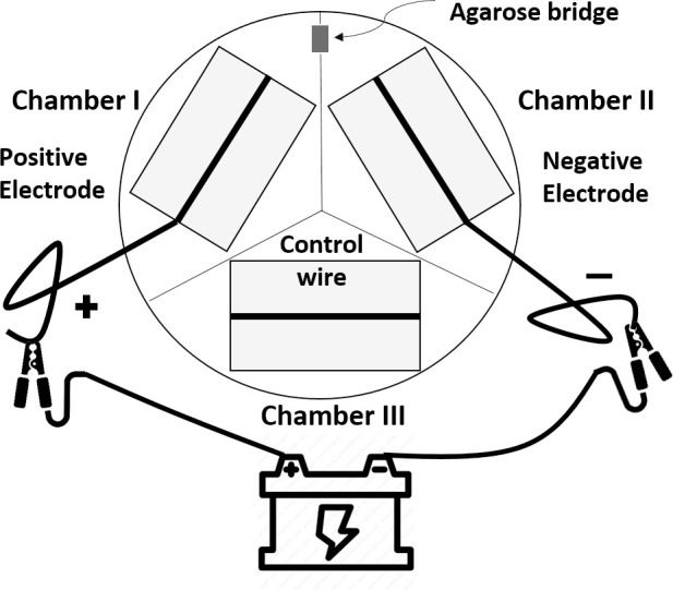
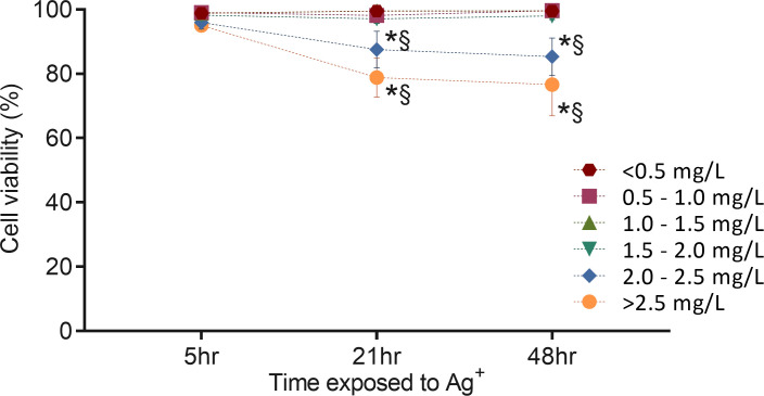
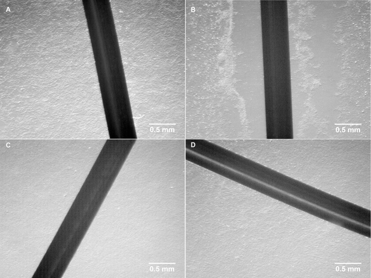
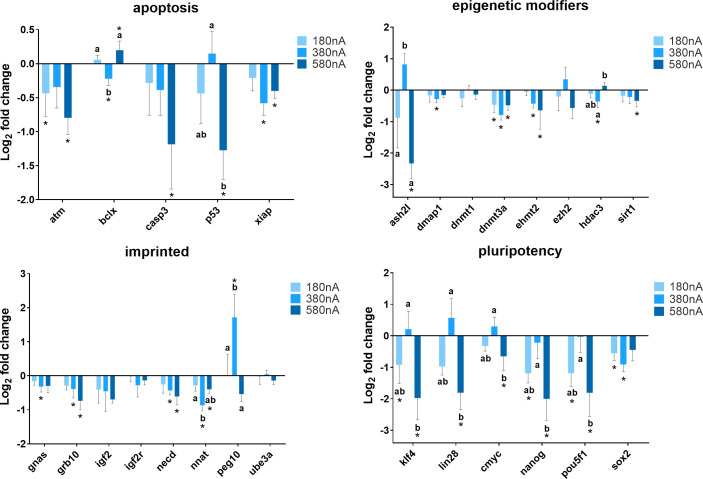
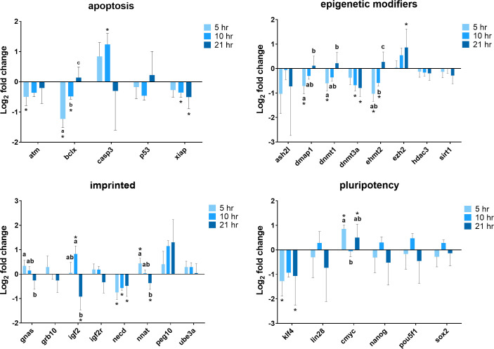
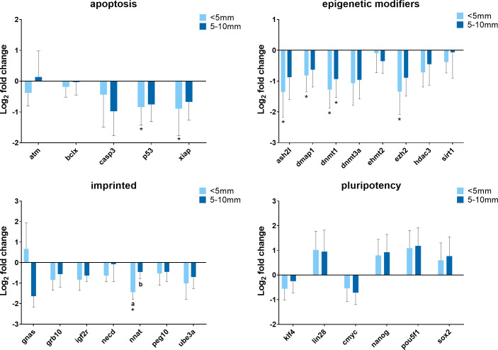
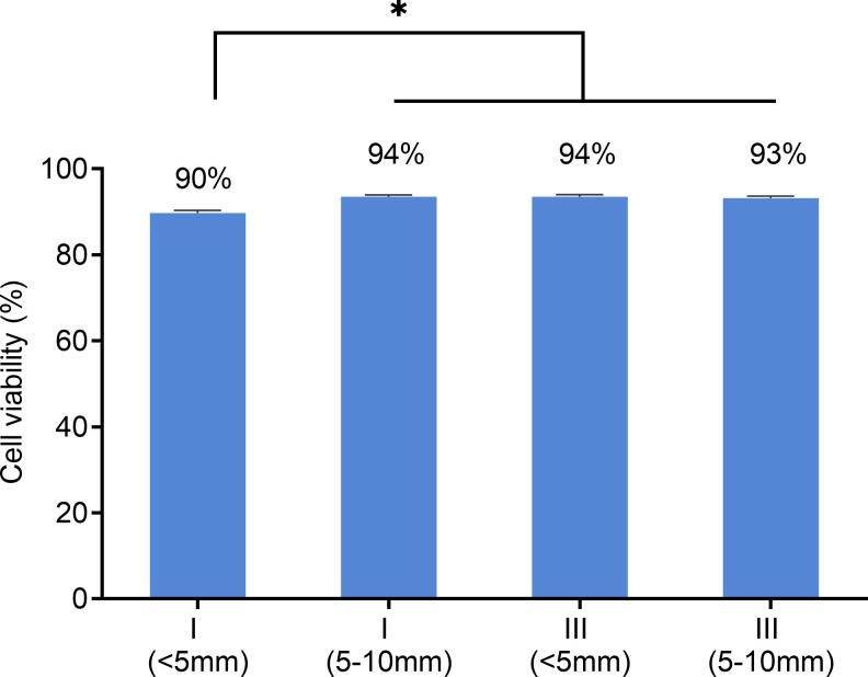
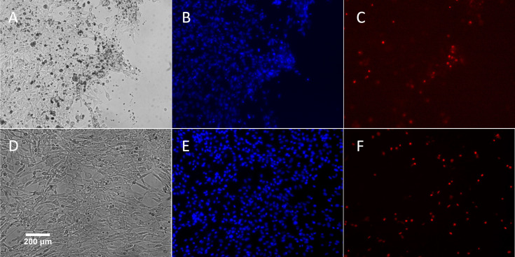
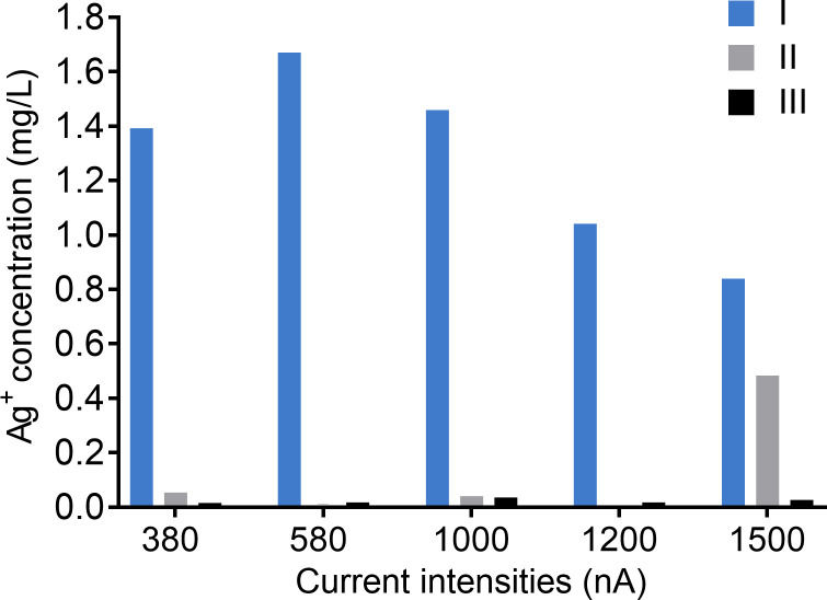
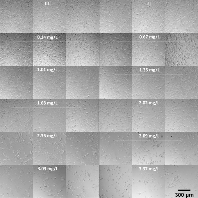
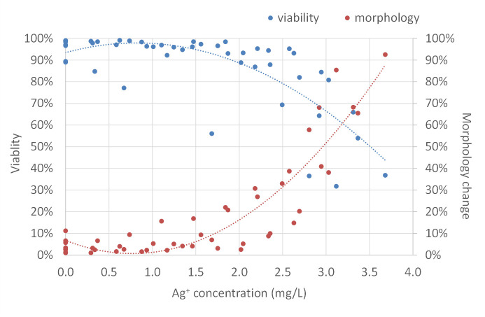
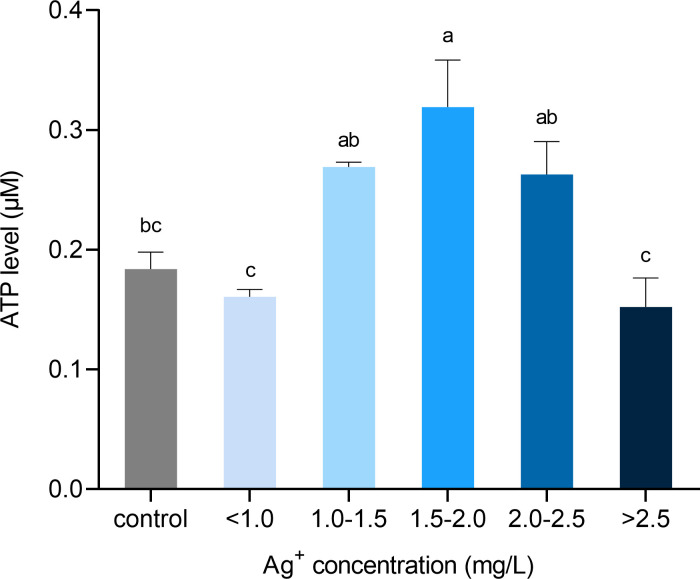
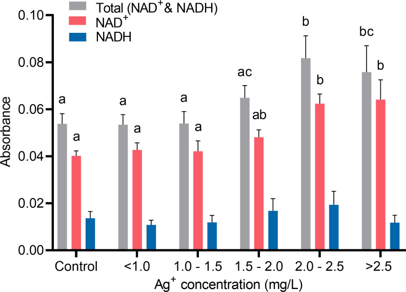
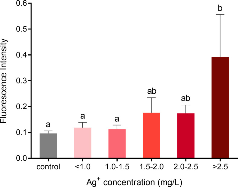
The medical applications of electrical biostimulation and silver ions have been evaluated in laboratory experiments and clinical studies for more than two decades. Their effects on preventing infection and promoting wound healing have been described. However, little is known about the role of electrical biostimulation and/or silver ion on changes in cellular transcriptome dynamics. To our knowledge, few studies have been conducted to investigate the potential of electrical biostimulation and silver ions in cell reprogramming. Besides, it is essential to assess any possible adverse effects or potential benefits of the silver ions on mammalian cells to address its safety concerns and to improve silver medical products. In this study, we investigated transcriptomic changes in porcine fibroblast cells in response to electrical biostimulation in the presence of silver ions. Exposed cells presented distinct morphological changes after treatment, which was mainly due to the exposure of silver ions rather than the electrical current itself. Gene expression analyses suggested that electrical biostimulation and silver ions did not increase the expression of pluripotency genes. Interestingly, a set of genes related to cellular metabolic processes were differentially expressed after cells were exposed to electrically generated silver ions for 21 hours. We found that 2.00 mg/L of electrically generated silver ion caused an increase of ATP generation and an increase of the total pool of NAD+ and NADH, while ROS production did not change. Aside from toxic effects, the results reported herein demonstrate the alternative effects of silver ions on mammalian cells, especially an oxidative phosphorylation burst. To our knowledge, this response of mammalian cells to silver ions has not been described previously. Although the function of this burst is not understood, it may lead to alterations in cellular activities such as metabolic remodeling and cell reprogramming, and/or serve an as-yet unknown function in neutralization or detoxification of the silver ions within the cells.

摘要

电生物刺激和银离子在医学中的应用已在实验室实验和临床研究中得到了二十多年的评估。它们在预防感染和促进伤口愈合方面的作用已被描述。然而,对于电生物刺激和/或银离子对细胞转录组动力学变化的影响知之甚少。据我们所知,很少有研究探讨电生物刺激和银离子在细胞重编程中的潜在作用。此外,评估银离子对哺乳动物细胞的任何潜在不良反应或潜在益处对于解决其安全性问题和改进银医疗产品至关重要。在这项研究中,我们研究了猪成纤维细胞在存在银离子的情况下对电生物刺激的转录组变化。处理后的细胞表现出明显的形态变化,这主要是由于银离子的暴露,而不是电流本身。基因表达分析表明,电生物刺激和银离子不会增加多能性基因的表达。有趣的是,一组与细胞代谢过程相关的基因在细胞暴露于电生成的银离子 21 小时后表达差异。我们发现,2.00mg/L 的电生成银离子会增加 ATP 的产生,并增加 NAD+和 NADH 的总池,而 ROS 的产生没有变化。除了毒性作用外,本文报告的结果还证明了银离子对哺乳动物细胞的替代作用,特别是氧化磷酸化爆发。据我们所知,哺乳动物细胞对银离子的这种反应以前尚未被描述过。虽然这种爆发的功能尚不清楚,但它可能导致细胞活动的改变,如代谢重塑和细胞重编程,以及/或在细胞内中和/或解毒银离子方面发挥未知的作用。

https://cdn.ncbi.nlm.nih.gov/pmc/blobs/1692/7875371/76283ff68cd6/pone.0246847.g014.jpg
https://cdn.ncbi.nlm.nih.gov/pmc/blobs/1692/7875371/b2dd3d89c575/pone.0246847.g001.jpg
https://cdn.ncbi.nlm.nih.gov/pmc/blobs/1692/7875371/b31dca17a0b7/pone.0246847.g002.jpg
https://cdn.ncbi.nlm.nih.gov/pmc/blobs/1692/7875371/aa4fa35f0452/pone.0246847.g003.jpg
https://cdn.ncbi.nlm.nih.gov/pmc/blobs/1692/7875371/6d9bd73d96b6/pone.0246847.g004.jpg
https://cdn.ncbi.nlm.nih.gov/pmc/blobs/1692/7875371/fa116874a448/pone.0246847.g005.jpg
https://cdn.ncbi.nlm.nih.gov/pmc/blobs/1692/7875371/da658872e8fb/pone.0246847.g006.jpg
https://cdn.ncbi.nlm.nih.gov/pmc/blobs/1692/7875371/4ac42177561c/pone.0246847.g007.jpg
https://cdn.ncbi.nlm.nih.gov/pmc/blobs/1692/7875371/04de2eef2a55/pone.0246847.g008.jpg
https://cdn.ncbi.nlm.nih.gov/pmc/blobs/1692/7875371/a341140ad544/pone.0246847.g009.jpg
https://cdn.ncbi.nlm.nih.gov/pmc/blobs/1692/7875371/cdb5c4396090/pone.0246847.g010.jpg
https://cdn.ncbi.nlm.nih.gov/pmc/blobs/1692/7875371/06f839f75c1d/pone.0246847.g011.jpg
https://cdn.ncbi.nlm.nih.gov/pmc/blobs/1692/7875371/c81f39d7717f/pone.0246847.g012.jpg
https://cdn.ncbi.nlm.nih.gov/pmc/blobs/1692/7875371/5c0322ab8d21/pone.0246847.g013.jpg
https://cdn.ncbi.nlm.nih.gov/pmc/blobs/1692/7875371/76283ff68cd6/pone.0246847.g014.jpg

相似文献

1
Effects of electrical biostimulation and silver ions on porcine fibroblast cells.电生物刺激和银离子对猪成纤维细胞的影响。
PLoS One. 2021 Feb 10;16(2):e0246847. doi: 10.1371/journal.pone.0246847. eCollection 2021.
2
Cytotoxicity and Transcriptomic Analysis of Silver Nanoparticles in Mouse Embryonic Fibroblast Cells.银纳米颗粒对小鼠胚胎成纤维细胞的细胞毒性及转录组学分析。
Int J Mol Sci. 2018 Nov 16;19(11):3618. doi: 10.3390/ijms19113618.
3
MicroRNA sequencing and molecular mechanisms analysis of the effects of gold nanoparticles on human dermal fibroblasts.金纳米颗粒对人真皮成纤维细胞影响的 miRNA 测序及分子机制分析。
Biomaterials. 2015 Jan;37:13-24. doi: 10.1016/j.biomaterials.2014.10.042. Epub 2014 Oct 22.
4
Silver nanoparticles induced neurotoxicity through oxidative stress in rat cerebral astrocytes is distinct from the effects of silver ions.银纳米颗粒通过氧化应激在大鼠脑星形胶质细胞中诱导的神经毒性不同于银离子的作用。
Neurotoxicology. 2016 Jan;52:210-21. doi: 10.1016/j.neuro.2015.09.007. Epub 2015 Dec 15.
5
Study of cytotoxicity mechanisms of silver nitrate in human dermal fibroblasts.硝酸银对人皮肤成纤维细胞细胞毒性机制的研究。
Toxicol Lett. 1998 Sep 15;98(3):169-79. doi: 10.1016/s0378-4274(98)00114-3.
6
Effect of biostimulation on oral fibroblast: a pilot study.生物刺激对口腔成纤维细胞的影响:一项初步研究。
J Biol Regul Homeost Agents. 2017 Dec 27;31(4 Suppl 2):139-145.
7
Increased NAD(P)H oxidase and reactive oxygen species in coronary arteries after balloon injury.球囊损伤后冠状动脉中NAD(P)H氧化酶和活性氧增加。
Arterioscler Thromb Vasc Biol. 2001 May;21(5):739-45. doi: 10.1161/01.atv.21.5.739.
8
Sub-cytotoxic concentrations of ionic silver promote the proliferation of human keratinocytes by inducing the production of reactive oxygen species.亚细胞毒性浓度的银离子通过诱导活性氧的产生来促进人角质形成细胞的增殖。
Front Med. 2018 Jun;12(3):289-300. doi: 10.1007/s11684-017-0550-7. Epub 2017 Nov 3.
9
SIRT3 mediates decrease of oxidative damage and prevention of ageing in porcine fetal fibroblasts.SIRT3介导猪胎儿成纤维细胞氧化损伤的降低及衰老的预防。
Life Sci. 2017 May 15;177:41-48. doi: 10.1016/j.lfs.2017.01.010. Epub 2017 Jan 26.
10
Comparison of different methods to study effects of silver nanoparticles on the pro- and antioxidant status of human keratinocytes and fibroblasts.比较不同方法以研究银纳米颗粒对人角质形成细胞和成纤维细胞的促氧化和抗氧化状态的影响。
Methods. 2016 Oct 15;109:55-63. doi: 10.1016/j.ymeth.2016.05.015. Epub 2016 May 20.

引用本文的文献

1
Electrical Stimulation of Cells: Drivers, Technology, and Effects.细胞的电刺激:驱动因素、技术及效应
Chem Rev. 2025 Aug 13;125(15):6874-6905. doi: 10.1021/acs.chemrev.4c00468. Epub 2025 Jul 17.
2
Bioelectronic Direct Current Stimulation at the Transition Between Reversible and Irreversible Charge Transfer.生物电子直流刺激在可逆和不可逆电荷转移之间的转变。
Adv Sci (Weinh). 2024 Jul;11(27):e2306244. doi: 10.1002/advs.202306244. Epub 2024 Mar 9.
3
Generation of direct current electrical fields as regenerative therapy for spinal cord injury: A review.

本文引用的文献

1
Antimicrobial Silver in Medicinal and Consumer Applications: A Patent Review of the Past Decade (2007⁻2017).药用及消费应用中的抗菌银:过去十年(2007 - 2017年)的专利综述
Antibiotics (Basel). 2018 Oct 26;7(4):93. doi: 10.3390/antibiotics7040093.
2
Revisiting the role of metabolism during development.重新探讨代谢在发育过程中的作用。
Development. 2018 Oct 1;145(19):dev131110. doi: 10.1242/dev.131110.
3
Combining electrical stimulation and tissue engineering to treat large bone defects in a rat model.电刺激与组织工程联合治疗大鼠模型中的大骨缺损。
产生直流电场作为脊髓损伤的再生疗法:综述
APL Bioeng. 2023 Sep 19;7(3):031505. doi: 10.1063/5.0152669. eCollection 2023 Sep.
4
Multiple Cell Cultures for MRI Analysis.多细胞培养物的 MRI 分析。
Int J Mol Sci. 2022 Sep 3;23(17):10109. doi: 10.3390/ijms231710109.
Sci Rep. 2018 Apr 20;8(1):6307. doi: 10.1038/s41598-018-24892-0.
4
Similarities and Differences between Silver Ions and Silver in Nanoforms as Antibacterial Agents.银离子和纳米形式银作为抗菌剂的异同。
Int J Mol Sci. 2018 Feb 2;19(2):444. doi: 10.3390/ijms19020444.
5
Toxicity of nano- and ionic silver to embryonic stem cells: a comparative toxicogenomic study.纳米银和离子银对胚胎干细胞的毒性:一项比较毒理基因组学研究。
J Nanobiotechnology. 2017 Apr 11;15(1):31. doi: 10.1186/s12951-017-0265-6.
6
Metabolic remodeling during the loss and acquisition of pluripotency.多能性丧失与获得过程中的代谢重塑。
Development. 2017 Feb 15;144(4):541-551. doi: 10.1242/dev.128389.
7
A review of reproductive and developmental toxicity of silver nanoparticles in laboratory animals.银纳米颗粒对实验动物生殖与发育毒性的综述。
Reprod Toxicol. 2017 Jan;67:149-164. doi: 10.1016/j.reprotox.2017.01.005. Epub 2017 Jan 11.
8
Mechanistic Basis of Antimicrobial Actions of Silver Nanoparticles.银纳米颗粒抗菌作用的机制基础
Front Microbiol. 2016 Nov 16;7:1831. doi: 10.3389/fmicb.2016.01831. eCollection 2016.
9
Differentially transcriptional regulation on cell cycle pathway by silver nanoparticles from ionic silver in larval zebrafish (Danio rerio).离子银中的银纳米颗粒对斑马鱼幼鱼细胞周期途径的差异转录调控。
Biochem Biophys Res Commun. 2016 Oct 28;479(4):753-758. doi: 10.1016/j.bbrc.2016.09.139. Epub 2016 Sep 28.
10
Evaluation of Silver Ion-Releasing Scaffolds in a 3D Coculture System of MRSA and Human Adipose-Derived Stem Cells for Their Potential Use in Treatment or Prevention of Osteomyelitis.在耐甲氧西林金黄色葡萄球菌(MRSA)与人类脂肪来源干细胞的三维共培养系统中评估银离子释放支架在治疗或预防骨髓炎方面的潜在用途。
Tissue Eng Part A. 2016 Nov;22(21-22):1258-1263. doi: 10.1089/ten.TEA.2016.0063. Epub 2016 Oct 25.