• 文献检索
  • 文档翻译
  • 深度研究
  • 学术资讯
  • Suppr Zotero 插件Zotero 插件
  • 邀请有礼
  • 套餐&价格
  • 历史记录
应用&插件
Suppr Zotero 插件Zotero 插件浏览器插件Mac 客户端Windows 客户端微信小程序
定价
高级版会员购买积分包购买API积分包
服务
文献检索文档翻译深度研究API 文档MCP 服务
关于我们
关于 Suppr公司介绍联系我们用户协议隐私条款
关注我们

Suppr 超能文献

核心技术专利:CN118964589B侵权必究
粤ICP备2023148730 号-1Suppr @ 2026

文献检索

告别复杂PubMed语法,用中文像聊天一样搜索,搜遍4000万医学文献。AI智能推荐,让科研检索更轻松。

立即免费搜索

文件翻译

保留排版,准确专业,支持PDF/Word/PPT等文件格式,支持 12+语言互译。

免费翻译文档

深度研究

AI帮你快速写综述,25分钟生成高质量综述,智能提取关键信息,辅助科研写作。

立即免费体验

CBCT 与口内成像评估种植体周围骨缺损的诊断准确性。

Diagnostic accuracy of CBCT versus intraoral imaging for assessment of peri-implant bone defects.

机构信息

OMFS IMPATH Research Group, Department of Imaging and Pathology, Faculty of Medicine, KU Leuven and Oral and Maxillofacial Surgery, University Hospitals Leuven, Kapucijnenvoer 33, 3000, Leuven, Belgium.

West China College of Stomatology, State Key Laboratory of Oral Disease & National Clinical Research Center for Oral Disease, Sichuan University, Chengdu, China.

出版信息

BMC Med Imaging. 2021 Feb 10;21(1):23. doi: 10.1186/s12880-021-00557-9.

DOI:10.1186/s12880-021-00557-9
PMID:33568085
原文链接:https://pmc.ncbi.nlm.nih.gov/articles/PMC7877020/
Abstract

BACKGROUND

Early detection of marginal bone loss is vital for treatment planning and prognosis of teeth and implant. This study was conducted to assess diagnostic accuracy of CBCT compared to intra-oral (IO) radiography for detection, classification, and measurement of peri-implant bone defects in an animal model.

METHODS

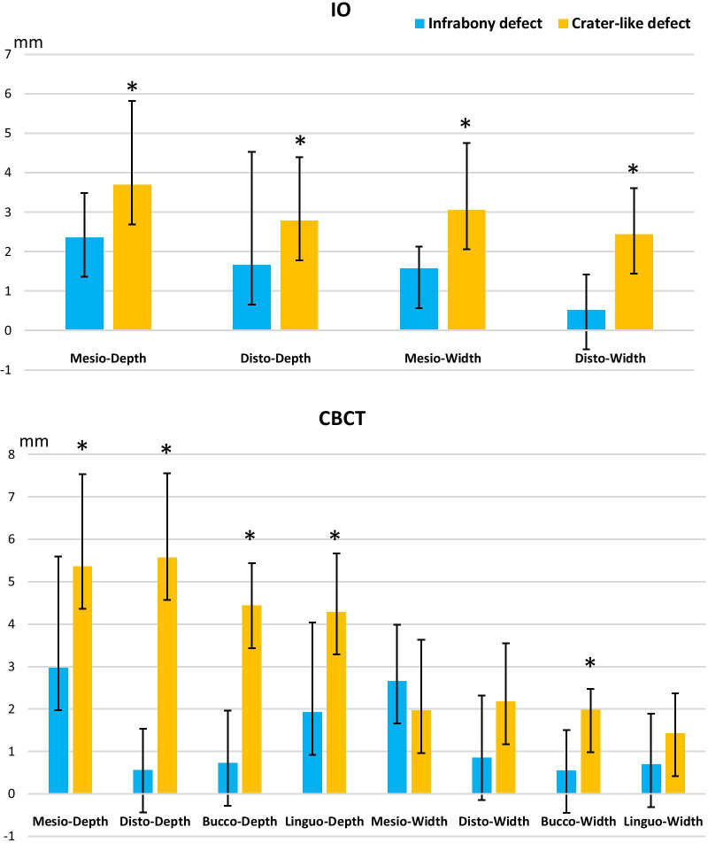
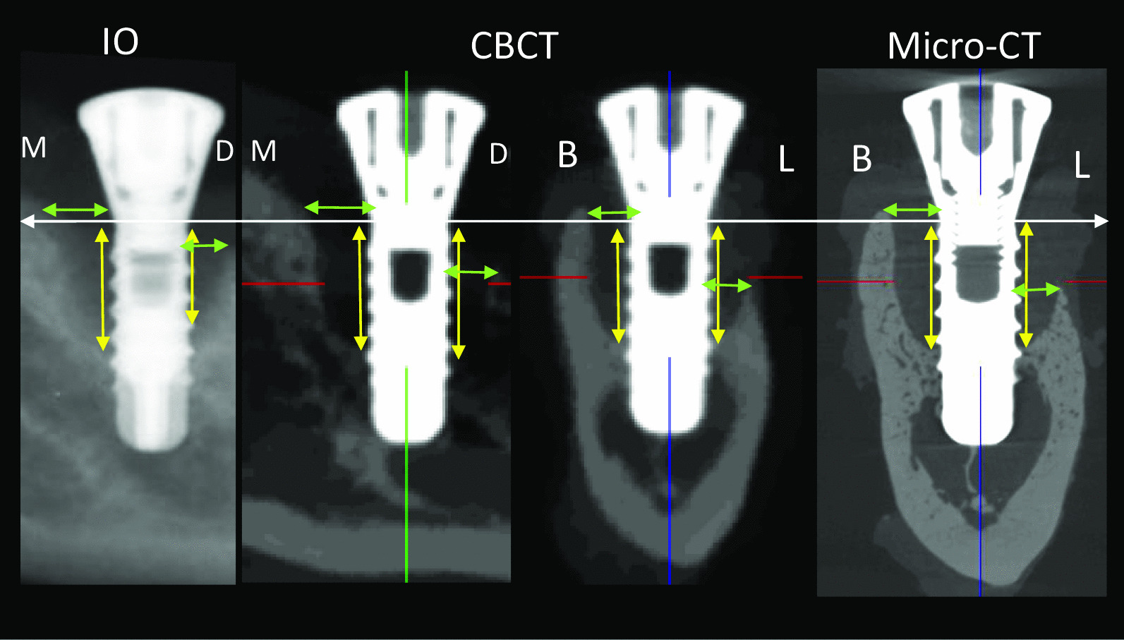
Fifty-four mandible blocks with implants were harvested from nine male health adult beagle dogs with acquisition of IO, CBCT and micro-CT images from all samples. Peri-implant bone defects from 16 samples were diagnosed using micro-CT and classified into 3 defect categories: dehiscence (n = 5), infrabony defect (n = 3) and crater-like defect (n = 8). Following training and calibration of the observers, they asked to detect location (mesial, distal, buccal, lingual) and shape of the defect (dehiscence, horizontal defect, vertical defect, carter-like defect) utilizing both IO and CBCT images. Both observers assessed defect depth and width on IO, CBCT and micro-CT images at each side of peri-implant bone defect via CT-analyzer software. Data were analyzed using SPSS software and a p value of < 0.05 was considered as statistically significant.

RESULTS

Overall, there was a high diagnostic accuracy for detection of bone defects with CBCT images (sensitivity: 100%/100%), while IO images showed a reduction in accuracy (sensitivity: 69%/63%). Similarly, diagnostic accuracy for defect classification was significantly higher for CBCT, whereas IO images were unable to correctly identify vestibular dehiscence, with incorrect assessment of half of the infrabony defects. For accuracy of measuring defect depth and width, a higher correlation was observed between CBCT and gold standard micro-CT (r = 0.91, 95% CI 0.86-0.94), whereas a lower correlation was seen for IO images (r = 0.82, 95% CI 0.67-0.91).

CONCLUSIONS

The diagnostic accuracy and reliability of CBCT was found to be superior to IO imaging for the detection, classification, and measurement of peri-implant bone defects. The application of CBCT adds substantial information related to the peri-implant bone defect diagnosis and decision-making which cannot be achieved with conventional IO imaging.

摘要

背景

早期发现边缘骨丢失对于牙齿和种植体的治疗计划和预后至关重要。本研究旨在评估 CBCT 与口腔内(IO)放射照相术相比,在动物模型中检测、分类和测量种植体周围骨缺损的诊断准确性。

方法

从 9 只健康成年比格犬的 54 个下颌骨块中采集样本,从所有样本中获取 IO、CBCT 和微 CT 图像。从 16 个样本中的种植体周围骨缺损使用微 CT 进行诊断,并将其分类为 3 种缺损类型:骨开裂(n=5)、骨内缺损(n=3)和火山口状缺损(n=8)。在对观察者进行培训和校准后,他们要求利用 IO 和 CBCT 图像来检测缺损的位置(近中、远中、颊侧、舌侧)和形状(骨开裂、水平缺损、垂直缺损、火山口状缺损)。两位观察者分别使用 CT 分析仪软件在 IO、CBCT 和微 CT 图像上评估每个种植体周围骨缺损侧的缺损深度和宽度。使用 SPSS 软件进行数据分析,p 值<0.05 被认为具有统计学意义。

结果

总体而言,CBCT 图像对骨缺损的检测具有很高的诊断准确性(敏感性:100%/100%),而 IO 图像的准确性则降低(敏感性:69%/63%)。同样,CBCT 对缺损分类的诊断准确性显著更高,而 IO 图像无法正确识别颊侧骨开裂,对一半的骨内缺损的评估不正确。对于测量缺损深度和宽度的准确性,CBCT 与金标准微 CT 之间的相关性更高(r=0.91,95%CI 0.86-0.94),而 IO 图像的相关性较低(r=0.82,95%CI 0.67-0.91)。

结论

与 IO 成像相比,CBCT 对种植体周围骨缺损的检测、分类和测量的诊断准确性和可靠性更高。CBCT 的应用增加了与种植体周围骨缺损诊断和决策相关的大量信息,而这是常规 IO 成像无法实现的。

https://cdn.ncbi.nlm.nih.gov/pmc/blobs/5330/7877020/e82fb4a1df89/12880_2021_557_Fig3_HTML.jpg
https://cdn.ncbi.nlm.nih.gov/pmc/blobs/5330/7877020/7598bfdedd00/12880_2021_557_Fig1_HTML.jpg
https://cdn.ncbi.nlm.nih.gov/pmc/blobs/5330/7877020/6985f264051f/12880_2021_557_Fig2_HTML.jpg
https://cdn.ncbi.nlm.nih.gov/pmc/blobs/5330/7877020/e82fb4a1df89/12880_2021_557_Fig3_HTML.jpg
https://cdn.ncbi.nlm.nih.gov/pmc/blobs/5330/7877020/7598bfdedd00/12880_2021_557_Fig1_HTML.jpg
https://cdn.ncbi.nlm.nih.gov/pmc/blobs/5330/7877020/6985f264051f/12880_2021_557_Fig2_HTML.jpg
https://cdn.ncbi.nlm.nih.gov/pmc/blobs/5330/7877020/e82fb4a1df89/12880_2021_557_Fig3_HTML.jpg

相似文献

1
Diagnostic accuracy of CBCT versus intraoral imaging for assessment of peri-implant bone defects.CBCT 与口内成像评估种植体周围骨缺损的诊断准确性。
BMC Med Imaging. 2021 Feb 10;21(1):23. doi: 10.1186/s12880-021-00557-9.
2
Accuracy of CBCT images in the assessment of buccal marginal alveolar peri-implant defects: effect of field of view.CBCT 图像评估颊侧边缘牙槽嵴种植体周围缺损的准确性:视野的影响。
Dentomaxillofac Radiol. 2014;43(4):20130332. doi: 10.1259/dmfr.20130332. Epub 2014 Mar 20.
3
Accuracy of cone-beam computed tomography, dental magnetic resonance imaging, and intraoral radiography for detecting peri-implant bone defects at single zirconia implants-An in vitro study.锥形束计算机断层扫描、口腔磁共振成像和口腔内射线照相术在检测单个氧化锆种植体周围骨缺损中的准确性——一项体外研究。
Clin Oral Implants Res. 2018 Sep;29(9):922-930. doi: 10.1111/clr.13348. Epub 2018 Aug 15.
4
Detection of peri-implant bone defects with different radiographic techniques - a human cadaver study.使用不同放射成像技术检测种植体周围骨缺损——一项人体尸体研究。
Clin Oral Implants Res. 2016 May;27(5):529-34. doi: 10.1111/clr.12619. Epub 2015 Jun 7.
5
Assessment of buccal marginal alveolar peri-implant and periodontal defects using a cone beam CT system with and without the application of metal artefact reduction mode.使用配备和不配备金属伪影降低模式的锥形束 CT 系统评估颊侧边缘牙槽嵴种植体和牙周缺损。
Dentomaxillofac Radiol. 2013;42(8):20130176. doi: 10.1259/dmfr.20130176.
6
Diagnostic Accuracy of Cone Beam Computed Tomography in Identifying Peri-implantitis-Like Bone Defects Ex Vivo.锥形束计算机断层扫描在体外识别种植体周围炎样骨缺损的诊断准确性。
Int J Periodontics Restorative Dent. 2021 Nov-Dec;41(6):e223-e231. doi: 10.11607/prd.5201.
7
Zirconia implants interfere with the evaluation of peri-implant bone defects in cone beam computed tomography (CBCT) images even with artifact reduction, a pilot study.氧化锆种植体即使采用了伪影减少技术,也会干扰 CBCT 图像中种植体周围骨缺损的评估:一项初步研究。
Dentomaxillofac Radiol. 2023 Nov;52(8):20230252. doi: 10.1259/dmfr.20230252. Epub 2023 Sep 4.
8
In vitro diagnostic accuracy of low-dose CBCT for evaluation of peri-implant bone lesions.低剂量 CBCT 评估种植体周围骨病变的体外诊断准确性。
Clin Oral Implants Res. 2019 Dec;30(12):1200-1208. doi: 10.1111/clr.13533. Epub 2019 Sep 20.
9
Accuracy of peri-implant bone evaluation using cone beam CT, digital intra-oral radiographs and histology.使用锥形束 CT、数字化口腔内放射影像和组织学评估种植体周围骨的准确性。
Dentomaxillofac Radiol. 2014;43(6):20130088. doi: 10.1259/dmfr.20130088. Epub 2014 May 2.
10
Assessment of peri-implant defects at titanium and zirconium dioxide implants by means of periapical radiographs and cone beam computed tomography: An in-vitro examination.通过根尖放射线片和锥形束计算机断层扫描评估钛和二氧化锆种植体周围缺损:一项体外研究。
Clin Oral Implants Res. 2018 Dec;29(12):1195-1201. doi: 10.1111/clr.13383. Epub 2018 Nov 22.

引用本文的文献

1
CBCT Validation Study on Intraclass Correlation for Linear Measurements in Peri-implantitis: an Observational Study.种植体周围炎线性测量组内相关系数的CBCT验证研究:一项观察性研究
J Oral Maxillofac Res. 2025 Mar 31;16(1):e4. doi: 10.5037/jomr.2025.16104. eCollection 2025 Jan-Mar.
2
Short-term cone beam CT study on bone mass changes post touch-controlled minimally invasive mandibular molar implant.触觉控制微创下颌磨牙种植术后骨量变化的短期锥形束CT研究
Sci Rep. 2025 Mar 29;15(1):10923. doi: 10.1038/s41598-024-84209-2.
3
Periapical radiographs vs cone beam CT imaging for the evaluation of peri-implant bone defects: an ex vivo study.

本文引用的文献

1
The ARRIVE guidelines 2.0: Updated guidelines for reporting animal research.ARRIVE 指南 2.0:报告动物研究的更新指南。
BMC Vet Res. 2020 Jul 14;16(1):242. doi: 10.1186/s12917-020-02451-y.
2
Effect of platelet-rich and platelet-poor plasma on peri-implant innervation in dog mandibles.富血小板血浆和贫血小板血浆对犬下颌骨种植体周围神经支配的影响。
Int J Implant Dent. 2019 Dec 4;5(1):40. doi: 10.1186/s40729-019-0193-3.
3
Effect of platelet-rich plasma on peri-implant trabecular bone volume and architecture: A preclinical micro-CT study in beagle dogs.
根尖片与锥形束CT成像在评估种植体周围骨缺损中的应用:一项体外研究
Med Oral Patol Oral Cir Bucal. 2025 May 1;30(3):e322-e332. doi: 10.4317/medoral.26777.
4
Challenges in detecting various peri-implant bone defects on modified intraoral oblique radiographic projections: evaluation of an artificial mandibular model.在改良口腔内斜位投照影像上检测各种种植体周围骨缺损的挑战:人工下颌骨模型的评估
Oral Radiol. 2025 Apr;41(2):248-259. doi: 10.1007/s11282-024-00798-8. Epub 2025 Jan 14.
5
A Split-Mouth Comparison Between Platelet-Rich Plasma and Lymphomyosot for Management of the Postoperative Sequelae After the Extraction of Impacted Third Molars: A Case Report.富血小板血浆与淋巴肌肉素治疗阻生第三磨牙拔除术后后遗症的双侧对照病例报告
Cureus. 2024 Nov 28;16(11):e74646. doi: 10.7759/cureus.74646. eCollection 2024 Nov.
6
Evaluation of the accuracy of cone beam computed tomography (CBCT) in the detection of peri-implant fenestration.评估锥形束计算机断层扫描(CBCT)在检测种植体周围开窗中的准确性。
BMC Oral Health. 2024 Aug 9;24(1):922. doi: 10.1186/s12903-024-04674-z.
7
Experimental models for peri-implant diseases: a narrative review.种植体周围疾病的实验模型:叙述性综述。
Clin Oral Investig. 2024 Jun 17;28(7):378. doi: 10.1007/s00784-024-05755-7.
8
Accuracy of implant height and width measurement with triaxial rotation method based on cone-beam CT.基于锥形束CT的三轴旋转法测量种植体高度和宽度的准确性
Heliyon. 2024 May 31;10(11):e32076. doi: 10.1016/j.heliyon.2024.e32076. eCollection 2024 Jun 15.
9
[Peri-implantitis: current concepts about its etiology, clinical and imaging characteristics. A review].[种植体周围炎:关于其病因、临床及影像学特征的当前概念。综述]
Rev Cient Odontol (Lima). 2023 Dec 26;10(4):e134. doi: 10.21142/2523-2754-1004-2022-134. eCollection 2023 Oct-Dec.
10
Influence of the Tube Angle on the Measurement Accuracy of Peri-Implant Bone Defects in Rectangular Intraoral X-ray Imaging.口腔内矩形X线成像中管角度对种植体周围骨缺损测量准确性的影响。
J Clin Med. 2024 Jan 10;13(2):391. doi: 10.3390/jcm13020391.
富血小板血浆对种植体周围小梁骨体积和结构的影响:一项在比格犬中的临床前 micro-CT 研究。
Clin Oral Implants Res. 2019 Dec;30(12):1190-1199. doi: 10.1111/clr.13532. Epub 2019 Sep 20.
4
Inaccuracy of buccal bone thickness estimation on cone-beam CT due to implant blooming: An ex-vivo study.基于锥形束 CT 的种植体伪影导致颊侧骨厚度估计不准确:一项离体研究。
J Clin Periodontol. 2019 Nov;46(11):1134-1143. doi: 10.1111/jcpe.13183. Epub 2019 Sep 9.
5
A controlled study on the accuracy and precision of intraoral radiography in assessing interproximal bone defect morphology around teeth and implants.一项关于口腔内放射摄影在评估牙齿和种植体周围邻间骨缺损形态的准确性和精确性的对照研究。
Eur J Oral Implantol. 2018;11(3):361-367.
6
Accuracy of cone-beam computed tomography, dental magnetic resonance imaging, and intraoral radiography for detecting peri-implant bone defects at single zirconia implants-An in vitro study.锥形束计算机断层扫描、口腔磁共振成像和口腔内射线照相术在检测单个氧化锆种植体周围骨缺损中的准确性——一项体外研究。
Clin Oral Implants Res. 2018 Sep;29(9):922-930. doi: 10.1111/clr.13348. Epub 2018 Aug 15.
7
Estimation of the diagnostic accuracy of clinical parameters for monitoring peri-implantitis progression: An experimental canine study.评估临床参数监测种植体周围炎进展的诊断准确性:一项实验性犬研究。
J Periodontol. 2018 Dec;89(12):1442-1451. doi: 10.1002/JPER.18-0081. Epub 2018 Aug 16.
8
Correlation of Three-Dimensional Radiologic Data with Subsequent Treatment Approach in Patients with Peri-implantitis: A Retrospective Analysis.种植体周围炎患者三维放射学数据与后续治疗方法的相关性:一项回顾性分析
Int J Periodontics Restorative Dent. 2017 Jul/Aug;37(4):481-489. doi: 10.11607/prd.2844.
9
Quantitative evaluation of metal artifacts using different CBCT devices, high-density materials and field of views.采用不同锥形束 CT 设备、高密度材料和视野定量评估金属伪影。
Clin Oral Implants Res. 2017 Dec;28(12):1509-1514. doi: 10.1111/clr.13019. Epub 2017 Apr 22.
10
Quantification of bone quality using different cone beam computed tomography devices: Accuracy assessment for edentulous human mandibles.使用不同锥形束计算机断层扫描设备对骨质量进行量化:无牙颌人类下颌骨的准确性评估。
Eur J Oral Implantol. 2016;9(4):411-424.