Department of Ophthalmology, The Second Affiliated Hospital of Jilin University, Changchun, 13000, China.
BMC Ophthalmol. 2021 Feb 10;21(1):78. doi: 10.1186/s12886-020-01785-3.
This research was conducted with the aim to determine the effect of diabetes mellitus on corneal endothelial cells.
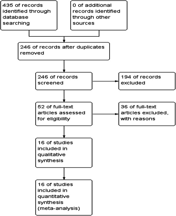
The terms: ("diabetes mellitus" or "diabetes" or "diabetic") and ("corneal endothelium" or "cornea" or "Corneas") searched in Pubmed, Embase, Cochrane, and Web of science until August 2019. The included types of studies contained observational studies. The standard mean difference (SMD) which was deemed as main size effects for continuous data was calculated by means and standard deviations. The data on corneal endothelial cell density (ECD), mean cell area (MCA), cell area variation coefficient (CV) and percentage of hexagonal cells (HEX) included in the study were collected and analyzed using stata15.1.
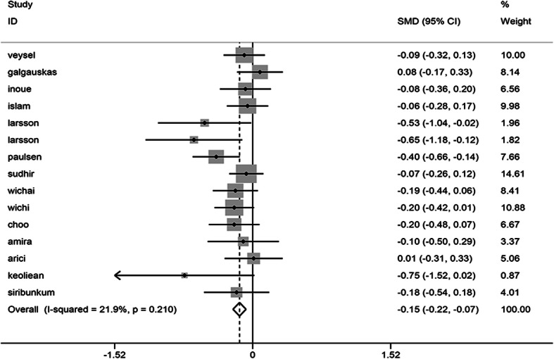
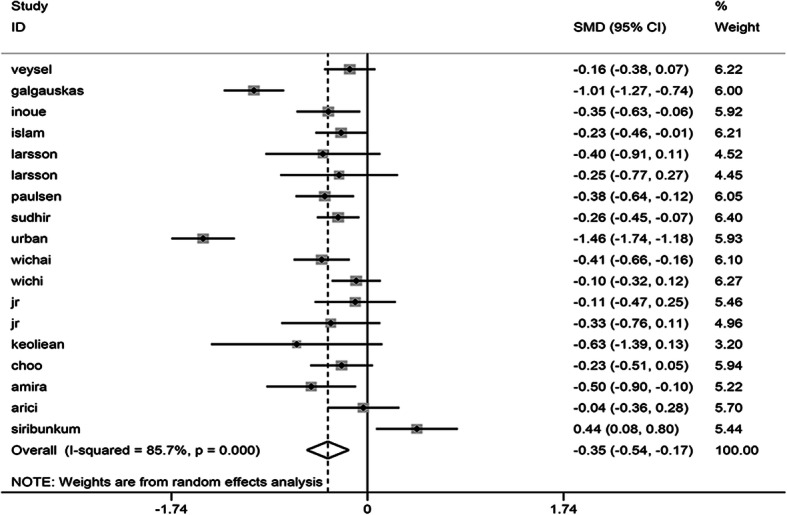
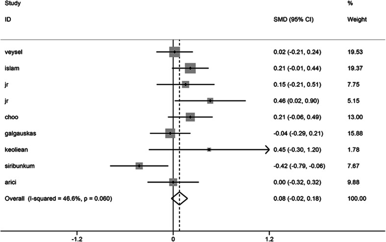
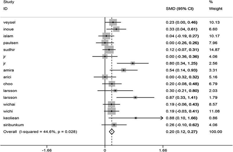
The final 16 cross-sectional studies and 2 case-control studies were included for the meta-analysis. Meta-analysis revealed that diabetes mellitus could reduce ECD (SMD = - 0.352, 95% CI -0.538, - 0.166) and the HEX (SMD = - 0.145, 95% CI -0.217, - 0.074), in addition to increasing CV (SMD = 0.195, 95% CI 0.123, 0.268). Nevertheless, there was no statistically significant differences observed when combining MCA (SMD = 0.078, 95% CI -0.022, 0.178). In subgroup analysis, Type 2 diabetes patients owned less corneal ECD (P < 0.05). Moreover the same results also found during the subgroup form Asia, Europe and American. The meta-regression revealed the type of diabetes mellitus might be contributing to heterogeneity. (P = 0.008). The results indicated a significant publication bias for studies, with combined CV (Begg's test, P = 0.006; Egger's test, P = 0.005) and merged combined HEX (Begg's test, P = 0.113; Egger's test, P = 0.024).
As indicated by meta-analysis, diabetes mellitus could cause a detrimental effect on corneal endothelium health. Diabetes mellitus contributed to the instability of corneal endothelium during the analysis. Therefore, further research is considered necessary to confirm our research results.
CED 42019145858 .
本研究旨在探讨糖尿病对角膜内皮细胞的影响。
在 Pubmed、Embase、Cochrane 和 Web of science 中检索“糖尿病”或“糖尿病”或“糖尿病患者”和“角膜内皮细胞”或“角膜”或“角膜”,检索时间截至 2019 年 8 月。纳入的研究类型包括观察性研究。采用均数和标准差计算主要大小效应的标准化均数差(SMD)。使用 stata15.1 收集和分析研究中包含的角膜内皮细胞密度(ECD)、平均细胞面积(MCA)、细胞面积变异系数(CV)和六边形细胞百分比(HEX)的数据。
最终纳入 16 项横断面研究和 2 项病例对照研究进行荟萃分析。Meta 分析显示,糖尿病可降低 ECD(SMD=-0.352,95%CI-0.538,-0.166)和 HEX(SMD=-0.145,95%CI-0.217,-0.074),并增加 CV(SMD=0.195,95%CI0.123,0.268)。然而,当合并 MCA(SMD=0.078,95%CI-0.022,0.178)时,差异无统计学意义。亚组分析显示,2 型糖尿病患者角膜 ECD 较少(P<0.05)。此外,在亚洲、欧洲和美洲的亚组中也发现了同样的结果。Meta 回归显示,糖尿病的类型可能导致异质性(P=0.008)。结果表明,研究存在显著的发表偏倚,合并 CV(Begg 检验,P=0.006;Egger 检验,P=0.005)和合并 HEX(Begg 检验,P=0.113;Egger 检验,P=0.024)。
Meta 分析表明,糖尿病可导致角膜内皮健康受损。糖尿病在分析过程中导致角膜内皮不稳定。因此,需要进一步研究以证实我们的研究结果。
CED 42019145858。