Hamza Mohamed N, Roshdy Maged Maher, Seleet Mouamen M, El Raggal Tamer M
Ophthalmology Department, Ain Shams University Hospitals, Cairo, Egypt.
Med Hypothesis Discov Innov Ophthalmol. 2021 Nov 17;10(3):121-128. doi: 10.51329/mehdiophthal1430. eCollection 2021 Fall.
To evaluate the normative values of corneal endothelial cell parameters within a group of healthy young Egyptian adults using specular microscopy and to examine any correlations between endothelial parameters and refractive or biometric parameters.
In this cross-sectional study, specular microscopy was used to study the right eyes of 150 healthy young volunteers and evaluated endothelial cell parameters, including cellular density, hexagonality (HEX), and coefficient of variation (CV) at 15 different points on the back corneal surface, which were later grouped into the central zone and either four quadrants or three annular zones. The same eyes underwent refractive and biometric assessments.
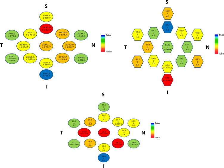
Hundred fifty healthy adults were examined, and the age ranged from 20 to 30 years, with a median of 23 (interquartile range, 21‒27) years. The mean (standard deviation) of central cell density was 2902.7 (270.7) cells/mm. The superior paracentral area had the lowest mean density (2895.8 cells/mm), but the highest mean HEX (67.7%), while the inferior peripheral area had the highest mean density (3100.5 cells/ mm) but the lowest mean HEX (64%). The difference in cell density among the three annular zones was not statistically significant ( = 0.365). However, HEX and CV in the central and paracentral zones differed statistically significantly from those of the peripheral zone ( < 0.001 and = 0.014, respectively). Weak but non-significant correlations were detected between endothelial cell density and all measured refractive and biometric parameters.
The findings of this study provided useful normative biometric and specular data in a specific age group and a specific population, and could be useful in planning intraocular surgery in young Egyptian adults. However, future longitudinal studies with a larger sample could refine more endothelial cell parameter specifications over time.
使用镜面显微镜评估一组健康的埃及年轻成年人角膜内皮细胞参数的正常数值,并检查内皮参数与屈光或生物测量参数之间的相关性。
在这项横断面研究中,使用镜面显微镜研究了150名健康年轻志愿者的右眼,并评估了角膜后表面15个不同点的内皮细胞参数,包括细胞密度、六边形性(HEX)和变异系数(CV),随后将这些点分为中央区以及四个象限或三个环形区。对同一只眼睛进行了屈光和生物测量评估。
检查了150名健康成年人,年龄范围为20至30岁,中位数为23(四分位间距,21 - 27)岁。中央细胞密度的平均值(标准差)为2902.7(270.7)个细胞/mm²。上旁中央区域的平均密度最低(2895.8个细胞/mm²),但平均HEX最高(67.7%),而下周边区域的平均密度最高(3100.5个细胞/mm²),但平均HEX最低(64%)。三个环形区之间的细胞密度差异无统计学意义(P = 0.365)。然而,中央区和旁中央区的HEX和CV与周边区相比有统计学显著差异(分别为P < 0.001和P = 0.014)。在内皮细胞密度与所有测量的屈光和生物测量参数之间检测到弱但无统计学意义的相关性。
本研究结果为特定年龄组和特定人群提供了有用的生物测量和镜面数据,可能有助于为埃及年轻成年人的眼内手术制定计划。然而,未来更大样本的纵向研究可能会随着时间的推移更精确地确定更多内皮细胞参数规范。