Department of Osteology and Biomechanics, University Medical Center Hamburg-Eppendorf, Hamburg, Germany.
Division of Orthopaedics, Department of Trauma and Orthopaedic Surgery, University Medical Center Hamburg-Eppendorf, Hamburg, Germany.
Osteoporos Int. 2021 Aug;32(8):1661-1668. doi: 10.1007/s00198-021-05881-y. Epub 2021 Feb 11.
We detected a high prevalence of low bone mineral density assessed by DXA in 268 elderly patients with end-stage osteoarthritis scheduled for total hip arthroplasty (18% osteoporosis, 41% osteopenia). Therefore, and due to the identified concomitant undertreatment, routine DXA measurements should be considered in elderly patients prior to surgery.
Bone quality represents a decisive factor for osseointegration, durability, and complications of an implanted prosthesis. Although the risk of osteoporosis increases with age and the assessment of bone mineral density (BMD) prior to total hip arthroplasty (THA) is recommended in elderly patients, a systematic, unbiased analysis of such patients is not available in the literature.
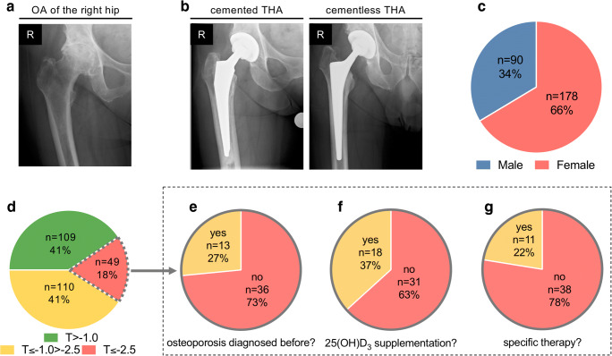
In this retrospective study, we examined 268 elderly patients (age ≥70 years) who underwent dual-energy X-ray absorptiometry (DXA) within 3 months prior to primary THA. Demographics, medical history, radiographic OA grade, and stem fixation method (i.e., cemented or cementless) were obtained.
In total, 153 (57%) cemented and 115 (43%) cementless stem fixations during THA were performed. Forty-nine patients (18%) were diagnosed with osteoporosis (T-score ≤-2.5), 110 patients (41%) with osteopenia (T-score ≤-1.0), and 109 patients (41%) with normal BMD (T-score >-1.0). Importantly, 36/49 patients (73%) with osteoporosis were not diagnosed before, resulting in a relevant undertreatment. Female sex and low body mass index (BMI) were the main factors negatively influencing the bone mineral density (BMD).
Due to a high incidence of undiagnosed and untreated osteoporosis in elderly patients with potential effects on the success of osseointegration as well as other clinical outcomes, DXA measurements should be included in the clinical routine for these patients prior to THA.
我们在 268 例行全髋关节置换术(THA)的终末期骨关节炎老年患者中发现 DXA 评估的低骨密度患病率较高(18%骨质疏松症,41%骨量减少)。因此,鉴于已确定的治疗不足,应在手术前对老年患者进行常规 DXA 测量。
骨质量是影响种植体骨整合、耐久性和并发症的决定性因素。尽管骨质疏松症的风险随着年龄的增长而增加,并且建议对老年患者进行 THA 前的骨密度(BMD)评估,但文献中尚无此类患者的系统、无偏分析。
在这项回顾性研究中,我们检查了 268 例行初次 THA 前 3 个月内进行双能 X 线吸收法(DXA)的老年患者(年龄≥70 岁)。获取了人口统计学资料、病史、放射学 OA 分级和股骨柄固定方法(即骨水泥固定或非骨水泥固定)。
共进行了 153 例(57%)骨水泥固定和 115 例(43%)非骨水泥固定的 THA。49 例(18%)患者诊断为骨质疏松症(T 评分≤-2.5),110 例(41%)为骨量减少症(T 评分≤-1.0),109 例(41%)为正常 BMD(T 评分>-1.0)。重要的是,骨质疏松症患者中有 36/49 例(73%)在术前未被诊断,导致治疗不足。女性和低体重指数(BMI)是对 BMD 有负面影响的主要因素。
由于老年患者骨质疏松症的发病率较高,且未得到诊断和治疗,这可能对骨整合的成功以及其他临床结果产生影响,因此,在 THA 前,应将 DXA 测量纳入这些患者的临床常规。