Electrical & Computer Engineering, Boston University, Boston, MA, USA.
Biomedical Engineering, Boston University, Boston, MA, USA.
Neoplasia. 2021 Mar;23(3):294-303. doi: 10.1016/j.neo.2021.01.005. Epub 2021 Feb 9.
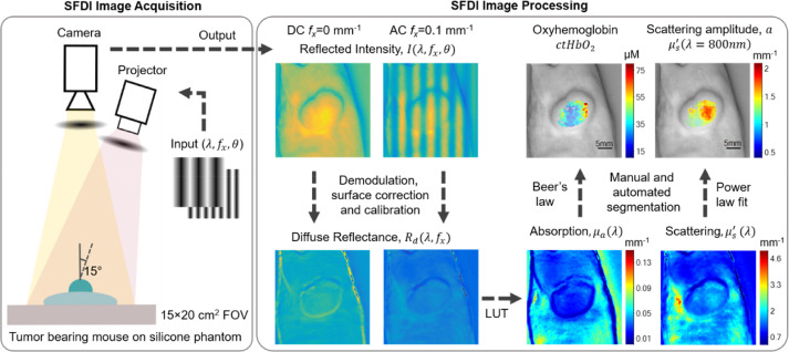
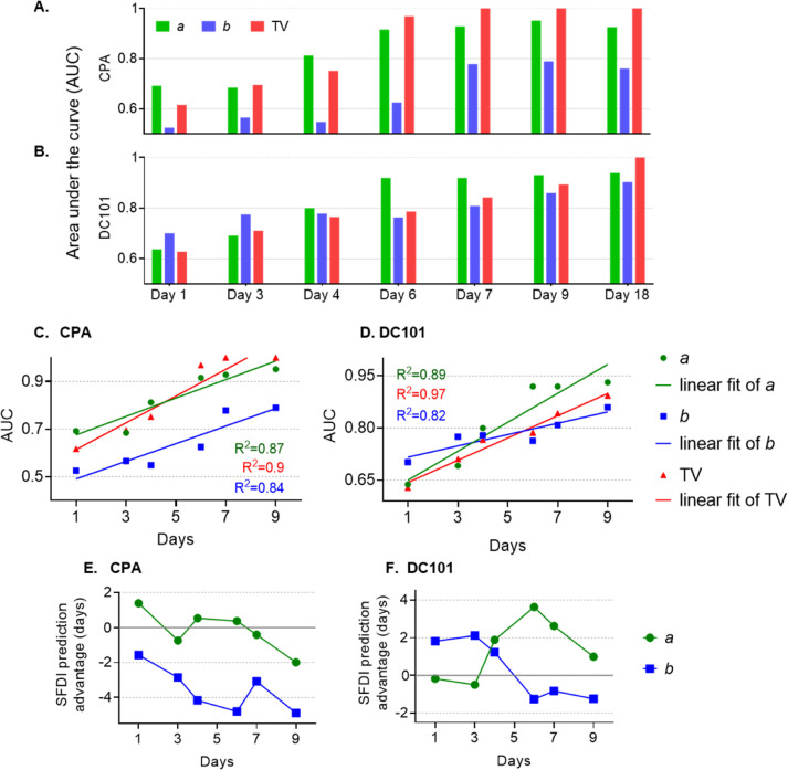
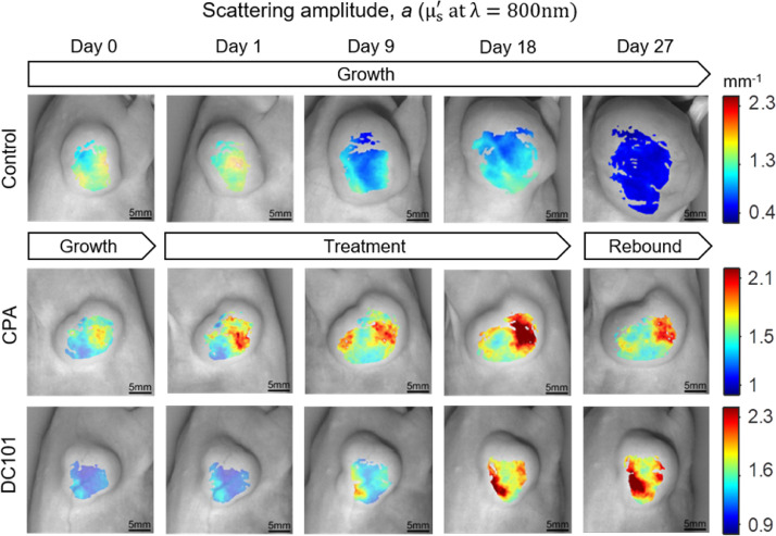
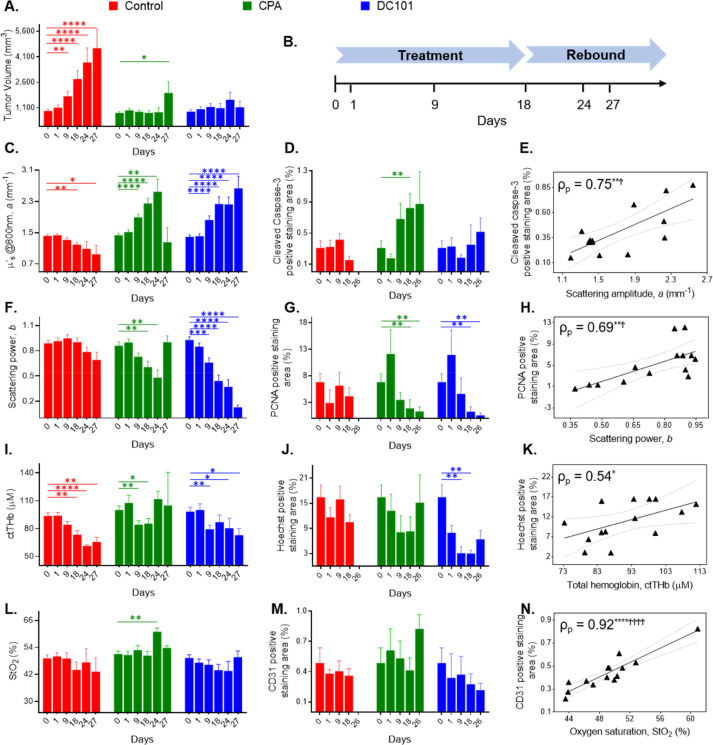
Monitoring of the in vivo tumor state to track therapeutic response in real time may help to evaluate new drug candidates, maximize treatment efficacy, and reduce the burden of overtreatment. Current preclinical tumor imaging methods have largely focused on anatomic imaging (e.g., MRI, ultrasound), functional imaging (e.g., FDG-PET), and molecular imaging with exogenous contrast agents (e.g., fluorescence optical tomography). Here we utalize spatial frequency domain imaging (SFDI), a noninvasive, label-free optical technique, for the wide-field quantification of changes in tissue optical scattering in preclinical tumor models during treatment with chemotherapy and antiangiogenic agents. Optical scattering is particularly sensitive to tissue micro-architectural changes, including those that occur during apoptosis, an early indicator of response to cytotoxicity induced by chemotherapy, thermotherapy, cryotherapy, or radiation therapy. We utilized SFDI to monitor responses of PC3/2G7 prostate tumors and E0771 mammary tumors to treatment with cyclophosphamide or the antiangiogenic agent DC101 for up to 49 days. The SFDI-derived scattering amplitude was highly correlated with cleaved caspase-3, a marker of apoptosis (ρ = 0.75), while the exponent of the scattering wavelength-dependence correlated with the cell proliferation marker PCNA (ρ = 0.69). These optical parameters outperformed tumor volume and several functional parameters (e.g., oxygen saturation and hemoglobin concentration) as an early predictive biomarker of treatment response. Quantitative diffuse optical scattering is thus a promising new early marker of treatment response, which does not require radiation or exogenous contrast agents.
实时监测体内肿瘤状态以跟踪治疗反应有助于评估新的候选药物,最大限度地提高治疗效果,并降低过度治疗的负担。目前的临床前肿瘤成像方法主要集中在解剖成像(例如 MRI、超声)、功能成像(例如 FDG-PET)和外源性对比剂的分子成像(例如荧光光学断层扫描)。在这里,我们利用空间域频域成像(SFDI),一种非侵入性、无标记的光学技术,对临床前肿瘤模型在化疗和抗血管生成剂治疗过程中组织光散射变化进行宽场定量。光散射对组织微观结构变化特别敏感,包括在化疗、热疗、冷冻疗法或放射疗法引起的细胞毒性诱导的细胞凋亡早期反应中发生的变化。我们利用 SFDI 监测 PC3/2G7 前列腺肿瘤和 E0771 乳腺肿瘤对环磷酰胺或抗血管生成剂 DC101 治疗的反应,最长可达 49 天。SFDI 衍生的散射幅度与细胞凋亡标志物 cleaved caspase-3 高度相关(ρ=0.75),而散射波长依赖性指数与细胞增殖标志物 PCNA 相关(ρ=0.69)。这些光学参数优于肿瘤体积和几种功能参数(例如,氧饱和度和血红蛋白浓度),是治疗反应的早期预测生物标志物。因此,定量漫射光散射是一种有前途的新的治疗反应早期标志物,它不需要辐射或外源性对比剂。