Department of Biomedical Engineering, University of Arkansas, Fayetteville, Arkansas.
Department of Biological Sciences, University of Arkansas, Fayetteville, Arkansas.
Radiat Res. 2022 Dec 1;198(6):545-552. doi: 10.1667/RADE-21-00228.1.
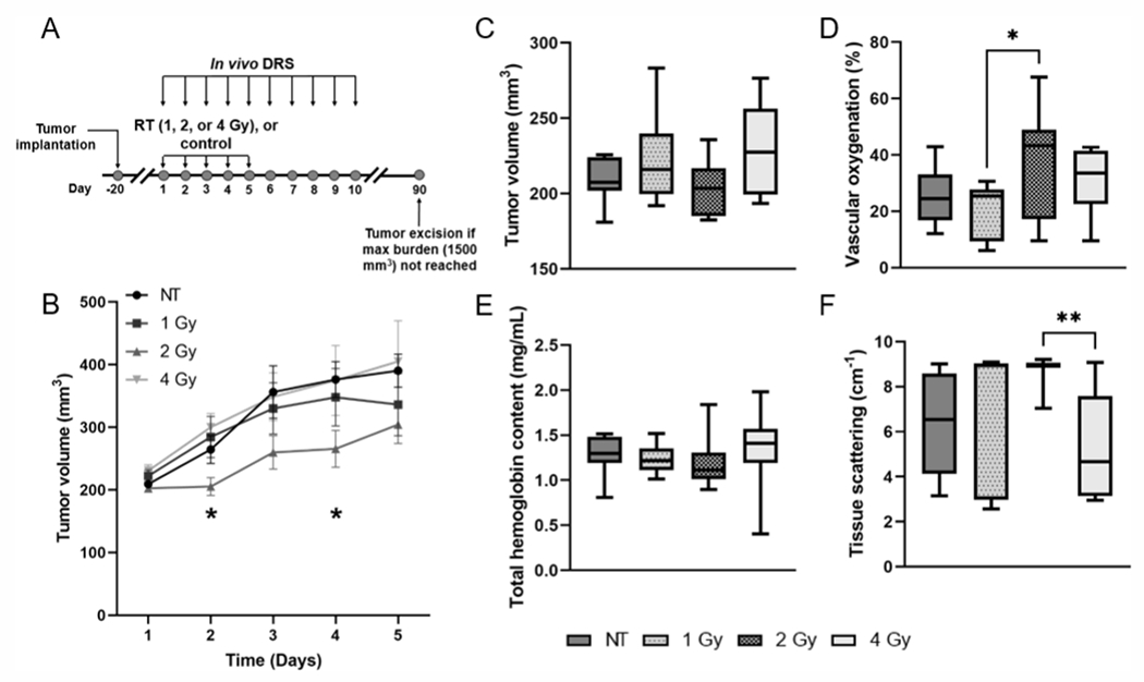
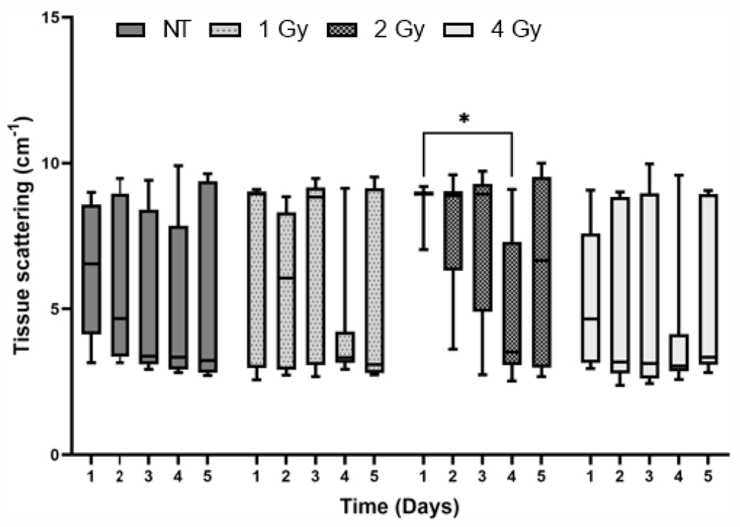
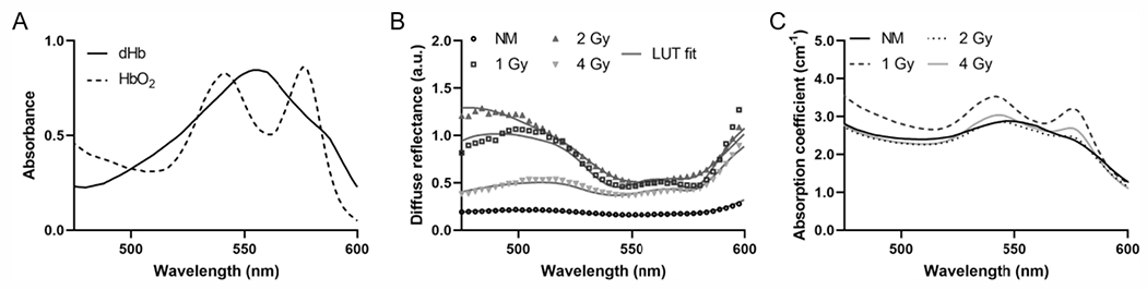
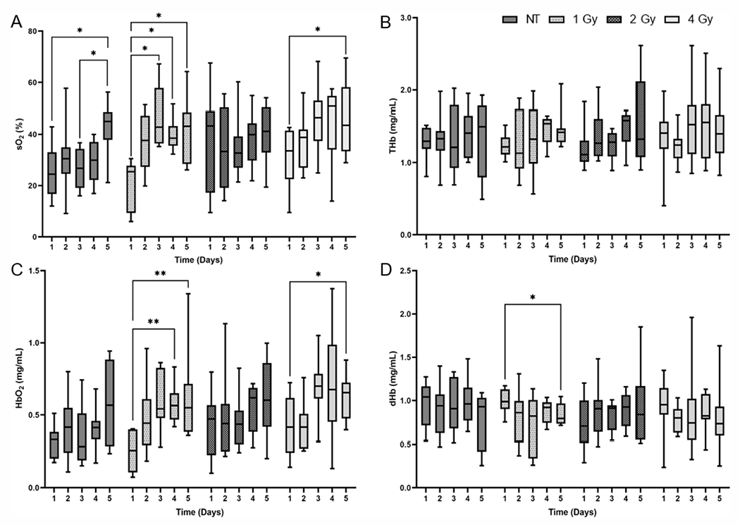
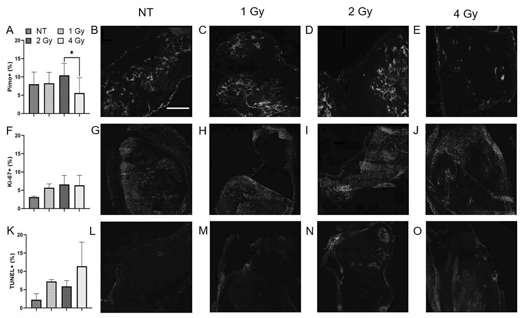
Radiation therapy plays an important role in cancer treatment, as it is an established method used as part of the treatment plan for the majority of cancer patients. Real-time monitoring of the effects of radiation on the tumor microenvironment can contribute to the development of better treatment plans. In this study, we use diffuse reflectance spectroscopy, a non-invasive optical fiber-based technique, to determine the effects of different doses of radiation on the tumor microenvironment, as well as to determine the sensitivity of diffuse reflectance spectroscopy to low doses of radiation that are used in the treatment of certain cancers. We injected 4T1 cells into 50 Balb/c mice to generate tumor xenografts. When the tumors grew to 200 mm3, we distributed the mice into a control group or one of three radiation groups: 1, 2, or 4 Gy/fraction, and they underwent treatment for five consecutive days. We measured the tumor volume and collected diffuse reflectance spectra every day, with optical measurements being acquired both before and one h postirradiation on the five days of treatment. Based on the diffusely reflected light, we quantified vascular oxygenation, total hemoglobin content, and tissue scattering within these tumors. There was a significant increase in tumor vascular oxygenation, which was primarily due to an increase in oxygenated hemoglobin, in response to a 1 Gy/fraction of radiation, while there was a decrease in tissue scattering in response to all doses of radiation. Immunohistochemical analysis revealed that tumor cell proliferation and apoptosis were higher in irradiated groups compared to the control group. Our findings show that diffuse reflectance spectroscopy is sensitive to microenvironmental changes in tumors treated with doses of radiation as low as 1 Gy/fraction.
放射治疗在癌症治疗中起着重要作用,因为它是一种既定的方法,被用于大多数癌症患者的治疗计划中。实时监测辐射对肿瘤微环境的影响有助于制定更好的治疗计划。在这项研究中,我们使用漫反射光谱学,一种非侵入性的光纤技术,来确定不同剂量的辐射对肿瘤微环境的影响,以及确定漫反射光谱学对用于治疗某些癌症的低剂量辐射的敏感性。我们将 4T1 细胞注射到 50 只 Balb/c 小鼠中,生成肿瘤异种移植物。当肿瘤生长到 200mm3 时,我们将小鼠分为对照组或三个放射组之一:1、2 或 4Gy/分次,连续治疗五天。我们测量肿瘤体积并每天收集漫反射光谱,在治疗的五天中,每天在辐照前后进行光学测量。基于漫反射光,我们定量了这些肿瘤内的血管氧合、总血红蛋白含量和组织散射。肿瘤血管氧合显著增加,这主要是由于 1Gy/分次的辐射导致氧合血红蛋白增加,而所有剂量的辐射都导致组织散射减少。免疫组织化学分析显示,与对照组相比,辐照组的肿瘤细胞增殖和凋亡更高。我们的研究结果表明,漫反射光谱学对低至 1Gy/分次的辐射剂量治疗的肿瘤微环境变化敏感。