Department of Vascular Surgery.
Department of Ultrasound.
Medicine (Baltimore). 2021 Feb 12;100(6):e24570. doi: 10.1097/MD.0000000000024570.
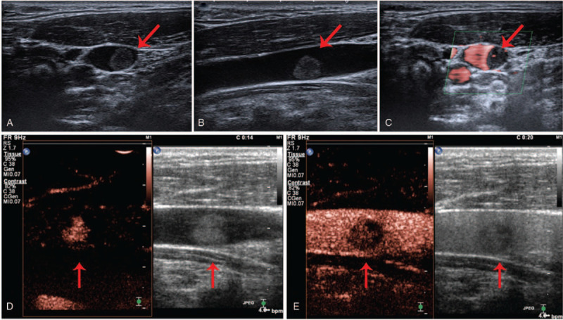
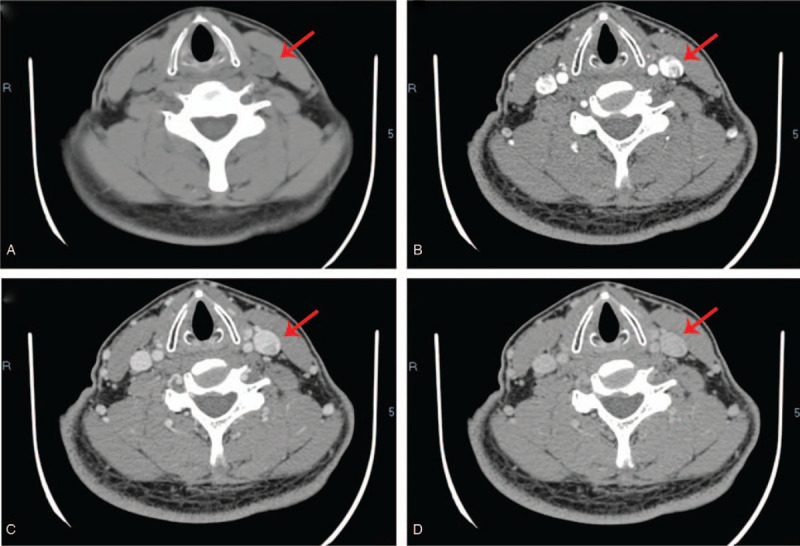
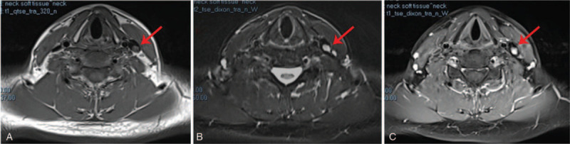
Intravenous pyogenic granuloma (IVPG) is a special type of pyogenic granuloma, and its preoperative diagnosis is difficult. We report a rare case of IVPG that develops in the lumen of the internal jugular vein (IJV). Here, we analyze the imaging characteristics of present case and summarize the imaging characteristics of previous reported cases.
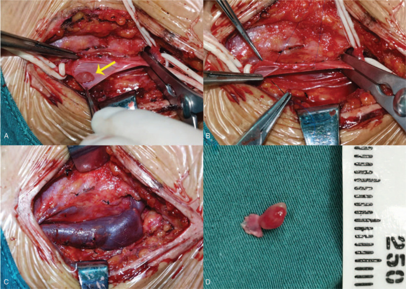
A 44-year-old man who presented with a growth in the IJV without any symptoms.
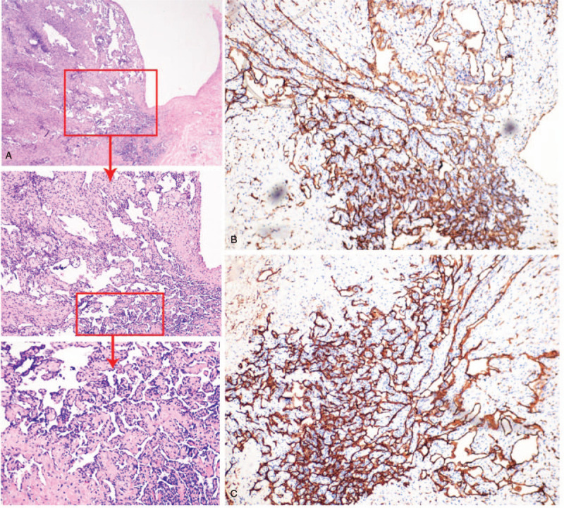
A diagnosis of IVPG was made, based on the pathological examination after surgery.
The patient underwent surgery to excise the vein segment containing the neoplasm.
The patient did not present with any complications in the postoperative follow-up period.
For clinician, IVPG's preoperative diagnosis is difficult. Although histopathology remains the gold standard for diagnosis, the combination of multiple types of imaging examinations is necessary to rule out the differential diagnoses of IVPG.
化脓性肉芽肿(PG)是一种特殊类型的化脓性肉芽肿,术前诊断困难。我们报告了一例罕见的发生于颈内静脉(IJV)管腔的 PG。在此,我们分析了本例的影像学特征,并总结了既往报道病例的影像学特征。
一名 44 岁男性,因 IJV 内生长物就诊,无任何症状。
基于术后病理检查,诊断为 IVPG。
患者接受了手术切除包含肿瘤的静脉段。
患者在术后随访期间未出现任何并发症。
对于临床医生来说,IVPG 的术前诊断较为困难。虽然组织病理学仍然是诊断的金标准,但需要结合多种类型的影像学检查以排除 IVPG 的鉴别诊断。