INSERM UEVE UMR861, I-STEM, AFM, Corbeil-Essonnes, France.
CECS, I-STEM, AFM, Corbeil-Essonnes, France.
J Cachexia Sarcopenia Muscle. 2021 Feb;12(1):209-232. doi: 10.1002/jcsm.12665. Epub 2021 Feb 14.
Duchenne muscular dystrophy (DMD) causes severe disability of children and death of young men, with an incidence of approximately 1/5000 male births. Symptoms appear in early childhood, with a diagnosis made mostly around 4 years old, a time where the amount of muscle damage is already significant, preventing early therapeutic interventions that could be more efficient at halting disease progression. In the meantime, the precise moment at which disease phenotypes arise-even asymptomatically-is still unknown. Thus, there is a critical need to better define DMD onset as well as its first manifestations, which could help identify early disease biomarkers and novel therapeutic targets.
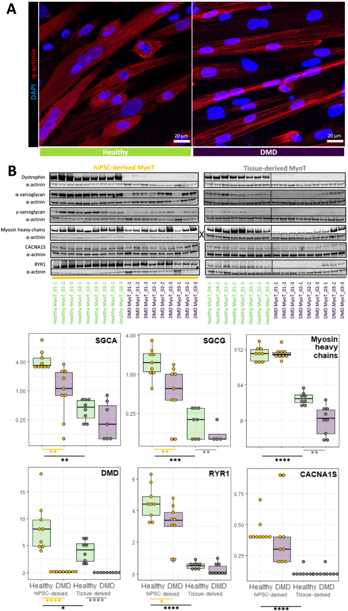
We have used both human tissue-derived myoblasts and human induced pluripotent stem cells (hiPSCs) from DMD patients to model skeletal myogenesis and compared their differentiation dynamics with that of healthy control cells by a comprehensive multi-omic analysis at seven time points. Results were strengthened with the analysis of isogenic CRISPR-edited human embryonic stem cells and through comparisons against published transcriptomic and proteomic datasets from human DMD muscles. The study was completed with DMD knockdown/rescue experiments in hiPSC-derived skeletal muscle progenitor cells and adenosine triphosphate measurement in hiPSC-derived myotubes.
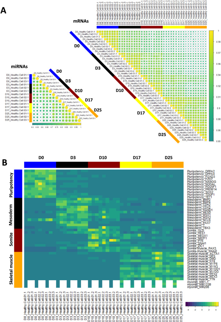
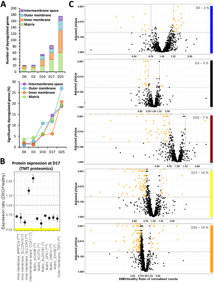
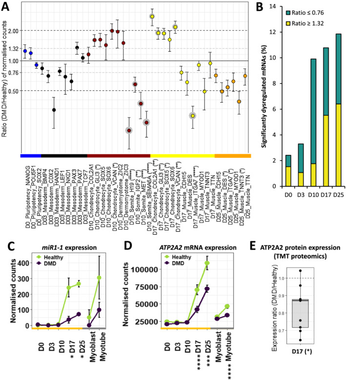
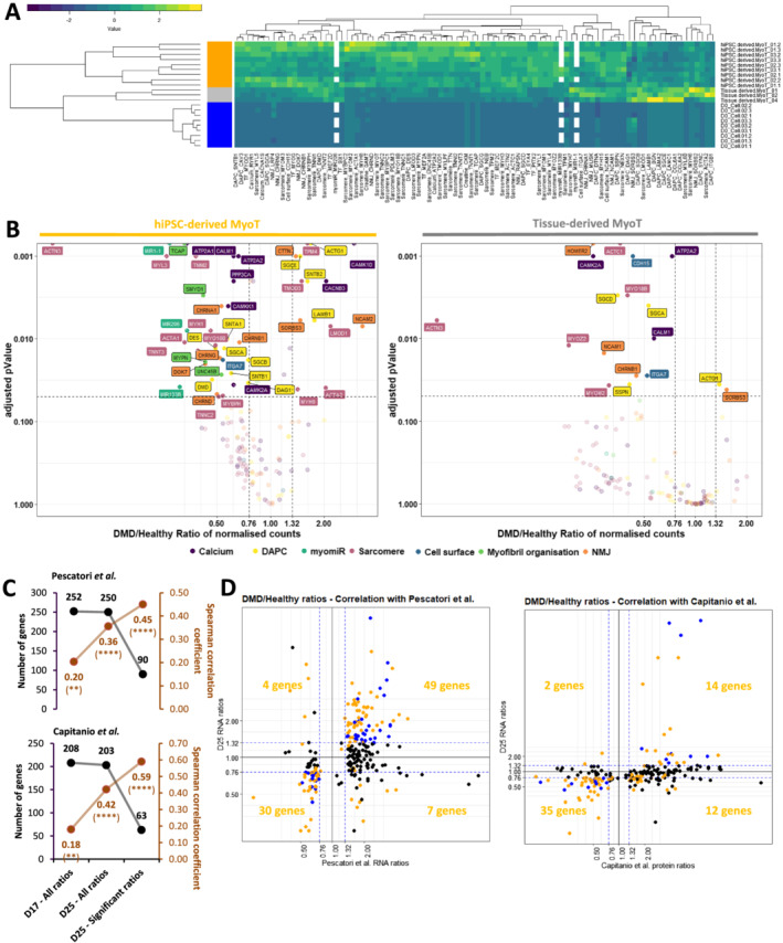
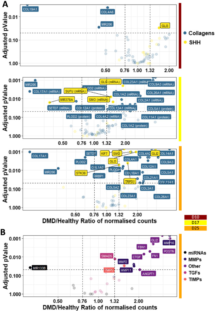
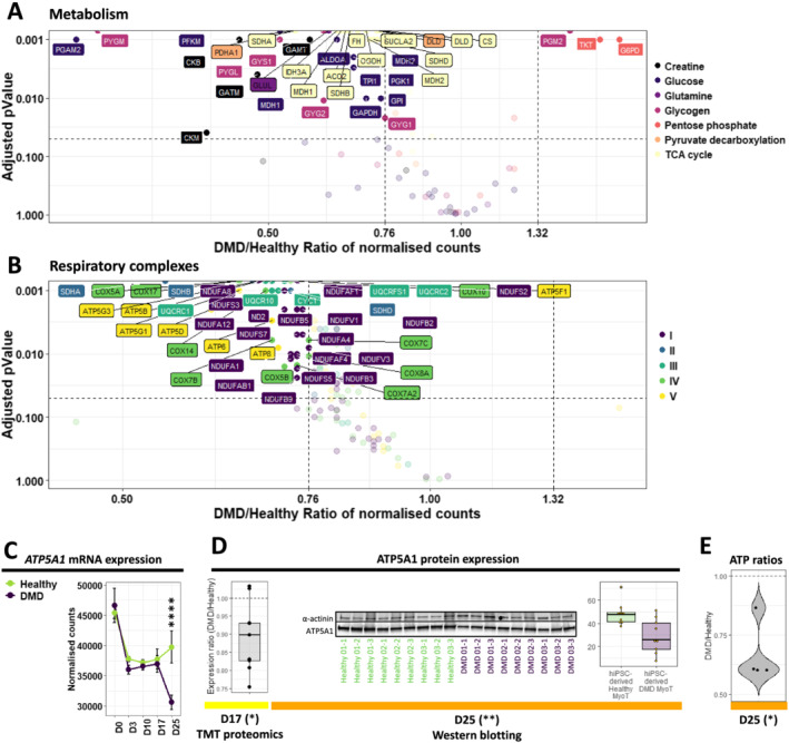
Transcriptome and miRnome comparisons combined with protein analyses demonstrated that hiPSC differentiation (i) leads to embryonic/foetal myotubes that mimic described DMD phenotypes at the differentiation endpoint and (ii) homogeneously and robustly recapitulates key developmental steps-mesoderm, somite, and skeletal muscle. Starting at the somite stage, DMD dysregulations concerned almost 10% of the transcriptome. These include mitochondrial genes whose dysregulations escalate during differentiation. We also describe fibrosis as an intrinsic feature of DMD skeletal muscle cells that begins early during myogenesis. All the omics data are available online for exploration through a graphical interface at https://muscle-dmd.omics.ovh/.
Our data argue for an early developmental manifestation of DMD whose onset is triggered before the entry into the skeletal muscle compartment, data leading to a necessary reconsideration of dystrophin roles during muscle development. This hiPSC model of skeletal muscle differentiation offers the possibility to explore these functions as well as find earlier DMD biomarkers and therapeutic targets.
杜氏肌营养不良症(DMD)导致儿童严重残疾和年轻男性死亡,发病率约为每 5000 名男性出生 1 例。症状出现在幼儿期,大多数诊断在 4 岁左右做出,此时肌肉损伤已经很严重,无法进行早期治疗干预,从而阻止疾病的进展。在此期间,疾病表型出现的准确时间——即使是无症状的——仍然未知。因此,迫切需要更好地定义 DMD 的发病时间及其最初表现,这有助于确定早期疾病生物标志物和新的治疗靶点。
我们使用人类组织衍生的成肌细胞和来自 DMD 患者的人类诱导多能干细胞(hiPSC)来模拟骨骼肌肉发生,并通过在七个时间点进行全面的多组学分析来比较它们与健康对照细胞的分化动态。通过对同源 CRISPR 编辑的人类胚胎干细胞的分析以及与人类 DMD 肌肉发表的转录组和蛋白质组数据集的比较,加强了结果。该研究还完成了 hiPSC 衍生的骨骼肌祖细胞中的 DMD 敲低/挽救实验和 hiPSC 衍生的肌管中的三磷酸腺苷测量。
转录组和 mirnome 比较结合蛋白质分析表明,hiPSC 分化(i)导致胚胎/胎儿肌管,在分化终点模拟描述的 DMD 表型,(ii)均匀且稳健地再现关键发育步骤——中胚层、体节和骨骼肌。从体节阶段开始,DMD 失调涉及近 10%的转录组。其中包括线粒体基因,其失调在分化过程中逐渐加剧。我们还描述了纤维化作为 DMD 骨骼肌细胞的内在特征,它在肌肉发生的早期开始。所有的组学数据都可在在线网站 https://muscle-dmd.omics.ovh/ 上通过图形界面进行探索。
我们的数据表明 DMD 的早期发育表现,其发病时间在进入骨骼肌之前就已经触发,这使得有必要重新考虑肌营养不良蛋白在肌肉发育中的作用。这种骨骼肌分化的 hiPSC 模型提供了探索这些功能以及寻找更早的 DMD 生物标志物和治疗靶点的可能性。