Anand Preeti, Pandey Jay Prakash, Pandey Dev Mani
Department of Bio-Engineering, Birla Institute of Technology, Mesra, Ranchi, Jharkhand, 835215, India.
Central Tasar Research and Training Institute, Piska- nagri, Jharkhand, Ranchi, India.
J Genet Eng Biotechnol. 2021 Feb 16;19(1):32. doi: 10.1186/s43141-021-00125-2.
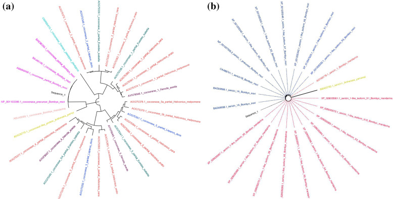
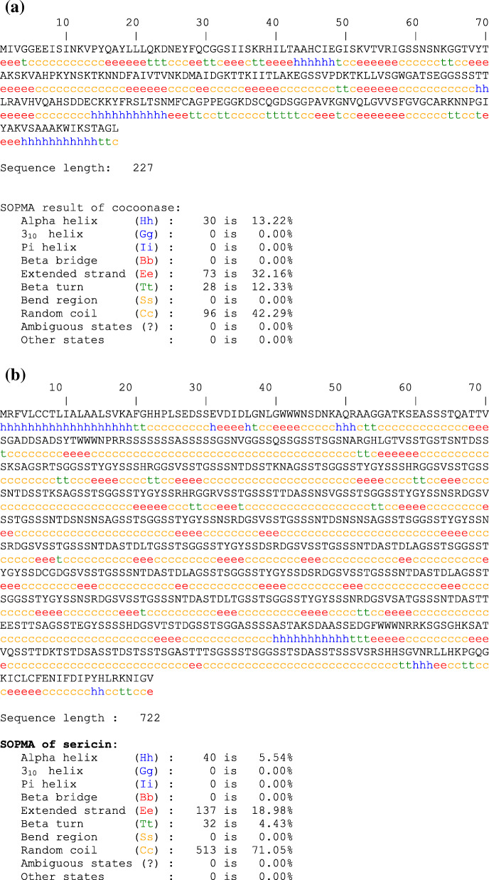
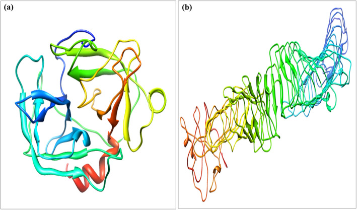
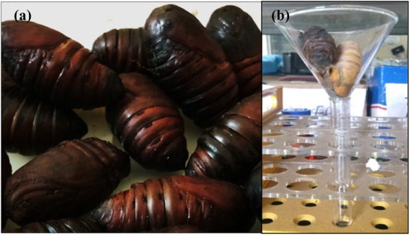
Cocoonase is a proteolytic enzyme that helps in dissolving the silk cocoon shell and exit of silk moth. Chemicals like anhydrous NaCO, Marseille soap, soda, ethylene diamine and tartaric acid-based degumming of silk cocoon shell have been in practice. During this process, solubility of sericin protein increased resulting in the release of sericin from the fibroin protein of the silk. However, this process diminishes natural color and softness of the silk. Cocoonase enzyme digests the sericin protein of silk at the anterior portion of the cocoon without disturbing the silk fibroin. However, no thorough characterization of cocoonase and sericin protein as well as imaging analysis of chemical- and enzyme-treated silk sheets has been carried out so far. Therefore, present study aimed for detailed characterization of cocoonase and sericin proteins, phylogenetic analysis, secondary and tertiary structure prediction, and computational validation as well as their interaction with other proteins. Further, identification of tasar silkworm (Antheraea mylitta) pupa stage for cocoonase collection, its purification and effect on silk sheet degumming, scanning electron microscope (SEM)-based comparison of chemical- and enzyme-treated cocoon sheets, and its optical coherence tomography (OCT)-based imaging analysis have been investigated. Various computational tools like Molecular Evolutionary Genetics Analysis (MEGA) X and Figtree, Iterative Threading Assembly Refinement (I-TASSER), self-optimized predicted method with alignment (SOPMA), PROCHECK, University of California, San Francisco (UCSF) Chimera, and Search Tool for the Retrieval of Interacting Genes/Proteins (STRING) were used for characterization of cocoonase and sericin proteins. Sodium dodecyl sulfate-polyacrylamide gel electrophoresis (SDS-PAGE), protein purification using Sephadex G 25-column, degumming of cocoon sheet using cocoonase enzyme and chemical NaCO, and SEM and OCT analysis of degummed cocoon sheet were performed.
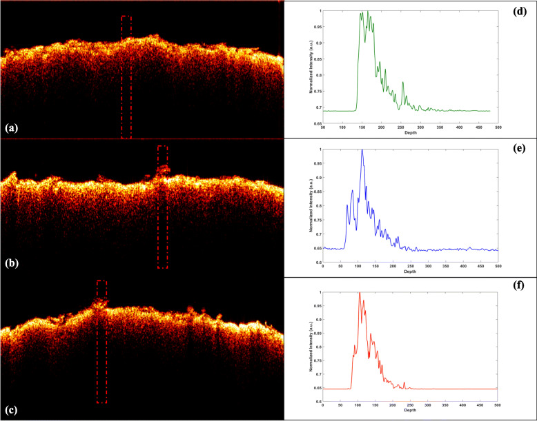
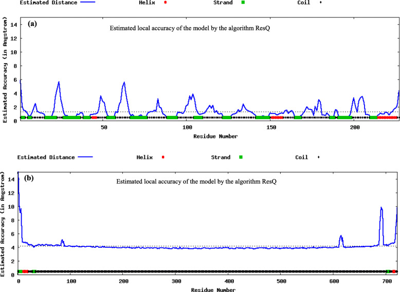
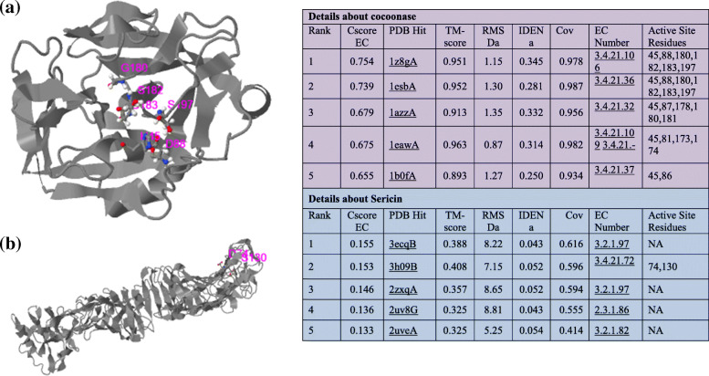
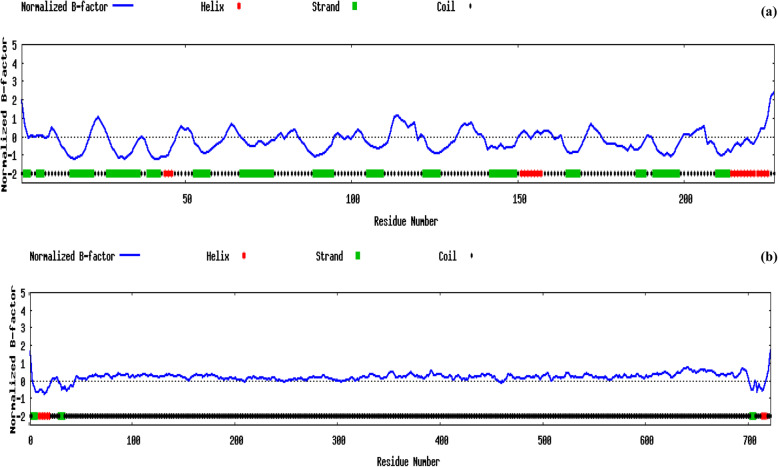
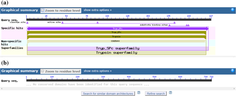
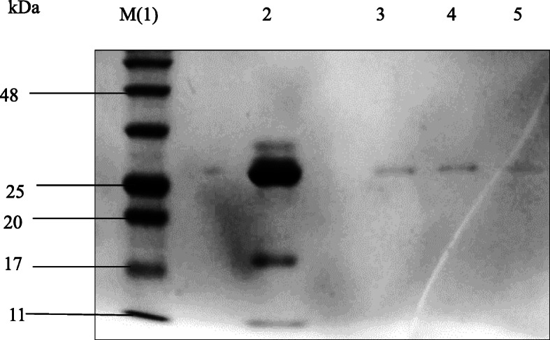
Predicted normalized B-factors of cocoonase and sericin with respect to α and β regions showed that these regions are structurally more stable in cocoonase while less stable in sericin. Conserved domain analysis revealed that B. mori cocoonase contains a trypsin-like serine protease with active site range 45 to 180 query sequences while substrate binding site from 175 to 200 query sequences. SDS-PAGE analysis of cocoonase indicated its molecular weight of 25-26 kDa. NaCO treatment showed more degumming effect (i.e., cocoon sheet weight loss) as compared to degumming with cocoonase. However, cocoonase-treated silk cocoon sheet holds the natural color of tasar silk, smoothness, and luster compared with the cocoon sheet treated with NaCO. SEM-based analysis showed the noticeable variation on the surface of silk fiber treated with cocoonase and NaCO. OCT analysis also exemplified the variations in the cross-sectional view of the cocoonase and NaCO-treated silk sheets.
Present study enlightens on the detailed characteristics of cocoonase and sericin proteins, comparative degumming activity, and image analysis of cocoonase enzyme and NaCO chemical-treated silk sheets. Obtained findings illustrated about use of cocoonase enzyme in the degumming of silk cocoon at larger scale that will be a boon to the silk industry.
茧酶是一种蛋白水解酶,有助于溶解蚕茧壳并使蚕蛾羽化。无水碳酸钠、马赛皂、苏打、乙二胺和基于酒石酸的蚕茧壳脱胶化学物质一直在实际应用中。在此过程中,丝胶蛋白的溶解度增加,导致丝胶从蚕丝的丝素蛋白中释放出来。然而,这个过程会降低丝绸的天然颜色和柔软度。茧酶能在不干扰丝素蛋白的情况下,消化蚕茧前部的丝胶蛋白。然而,到目前为止,尚未对茧酶和丝胶蛋白进行全面表征,也未对化学处理和酶处理的丝绸片进行成像分析。因此,本研究旨在对茧酶和丝胶蛋白进行详细表征、系统发育分析、二级和三级结构预测以及计算验证,以及它们与其他蛋白质的相互作用。此外,还研究了用于收集茧酶的柞蚕蛹期的鉴定、其纯化及其对丝绸片脱胶的影响、基于扫描电子显微镜(SEM)的化学处理和酶处理蚕茧片的比较,以及基于光学相干断层扫描(OCT)的成像分析。使用了多种计算工具,如分子进化遗传学分析(MEGA)X和Figtree、迭代穿线装配优化(I-TASSER)、带比对的自优化预测方法(SOPMA)、PROCHECK、加利福尼亚大学旧金山分校(UCSF)Chimera以及用于检索相互作用基因/蛋白质的搜索工具(STRING)来表征茧酶和丝胶蛋白。进行了十二烷基硫酸钠-聚丙烯酰胺凝胶电泳(SDS-PAGE)、使用葡聚糖G 25柱进行蛋白质纯化、使用茧酶和化学物质碳酸钠对蚕茧片进行脱胶,以及对脱胶蚕茧片进行SEM和OCT分析。
相对于α和β区域,茧酶和丝胶的预测归一化B因子表明,这些区域在茧酶中结构更稳定,而在丝胶中稳定性较差。保守结构域分析表明,家蚕茧酶含有一种胰蛋白酶样丝氨酸蛋白酶,其活性位点范围为45至180个查询序列,而底物结合位点为175至200个查询序列。茧酶的SDS-PAGE分析表明其分子量为25 - 26 kDa。与用茧酶脱胶相比,碳酸钠处理显示出更大的脱胶效果(即蚕茧片重量损失)。然而,与用碳酸钠处理的蚕茧片相比,用茧酶处理的蚕茧片保留了柞蚕丝的天然颜色、光滑度和光泽。基于SEM的分析显示了用茧酶和碳酸钠处理的丝纤维表面的显著差异。OCT分析也例证了茧酶和碳酸钠处理的丝绸片横截面视图的差异。
本研究揭示了茧酶和丝胶蛋白的详细特征、比较脱胶活性以及茧酶和碳酸钠化学处理的丝绸片的图像分析。所得结果表明,茧酶在大规模蚕茧脱胶中的应用将对丝绸行业有益。