Medical Imaging Department, First Affiliated Hospital of Kunming Medical University, Kunming, 650000, China.
Precision Health Institution, PDx, GE Healthcare (China), Beijing, 100176, China.
BMC Med Imaging. 2021 Feb 17;21(1):31. doi: 10.1186/s12880-021-00564-w.
In this COVID-19 pandemic, the differential diagnosis of viral pneumonia is still challenging. We aimed to assess the classification performance of computed tomography (CT)-based CT signs and radiomics features for discriminating COVID-19 and influenza pneumonia.
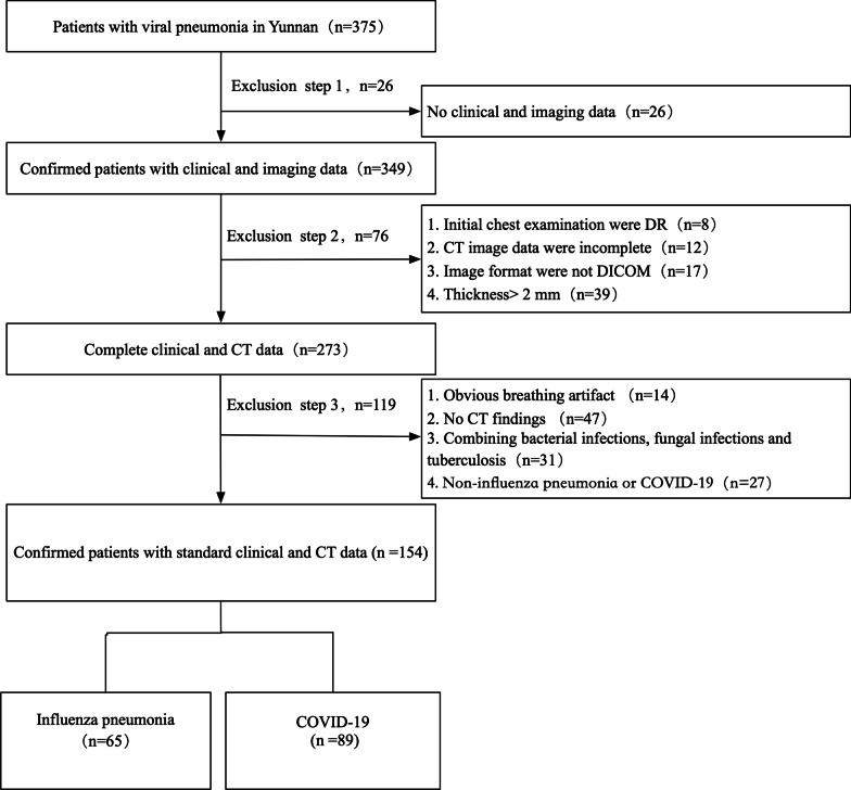
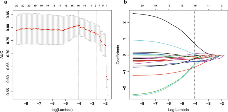
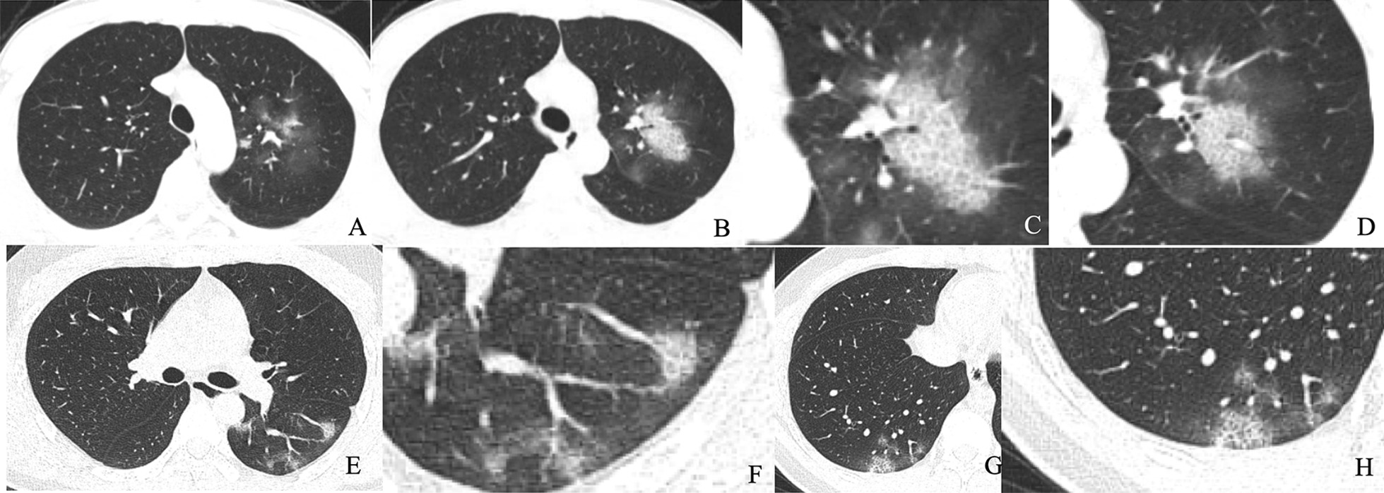
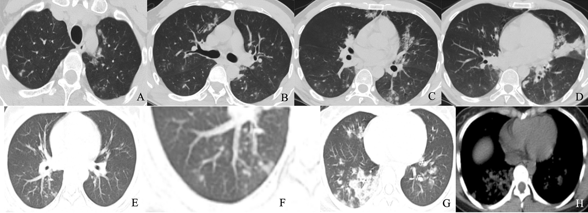
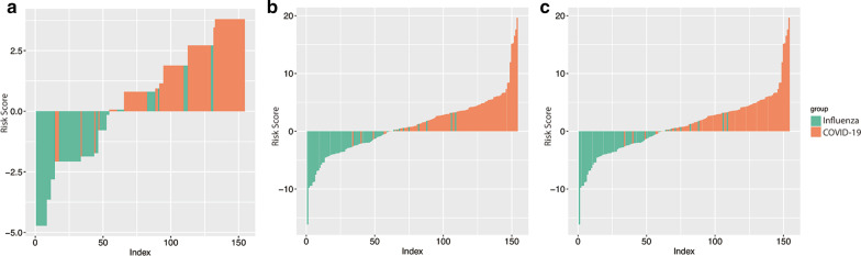
A total of 154 patients with confirmed viral pneumonia (COVID-19: 89 cases, influenza pneumonia: 65 cases) were collected retrospectively in this study. Pneumonia signs and radiomics features were extracted from the initial unenhanced chest CT images to build independent and combined models. The predictive performance of the radiomics model, CT sign model, the combined model was constructed based on the whole dataset and internally invalidated by using 1000-times bootstrap. Diagnostic performance of the models was assessed via receiver operating characteristic (ROC) analysis.
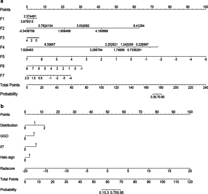
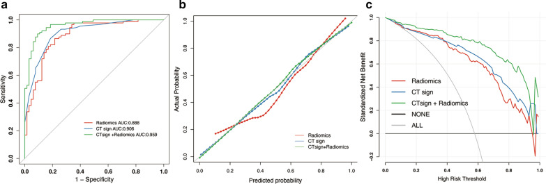
The combined models consisted of 4 significant CT signs and 7 selected features and demonstrated better discrimination performance between COVID-19 and influenza pneumonia than the single radiomics model. For the radiomics model, the area under the ROC curve (AUC) was 0.888 (sensitivity, 86.5%; specificity, 78.4%; accuracy, 83.1%), and the AUC was 0.906 (sensitivity, 86.5%; specificity, 81.5%; accuracy, 84.4%) in the CT signs model. After combining CT signs and radiomics features, AUC of the combined model was 0.959 (sensitivity, 89.9%; specificity, 90.7%; accuracy, 90.3%).
CT-based radiomics combined with signs might be a potential method for distinguishing COVID-19 and influenza pneumonia with satisfactory performance.
在本次 COVID-19 大流行中,病毒性肺炎的鉴别诊断仍然具有挑战性。我们旨在评估基于计算机断层扫描(CT)的 CT 征象和放射组学特征对鉴别 COVID-19 和流感肺炎的分类性能。
本研究回顾性收集了 154 例确诊为病毒性肺炎的患者(COVID-19:89 例,流感肺炎:65 例)。从初始未增强胸部 CT 图像中提取肺炎征象和放射组学特征,以建立独立和联合模型。基于全数据集构建放射组学模型、CT 征象模型和联合模型,并使用 1000 次自举法进行内部验证。使用受试者工作特征(ROC)分析评估模型的诊断性能。
联合模型由 4 个显著的 CT 征象和 7 个选定的特征组成,与单一放射组学模型相比,对 COVID-19 和流感肺炎的鉴别性能更好。对于放射组学模型,ROC 曲线下面积(AUC)为 0.888(敏感性为 86.5%,特异性为 78.4%,准确性为 83.1%),CT 征象模型的 AUC 为 0.906(敏感性为 86.5%,特异性为 81.5%,准确性为 84.4%)。在联合 CT 征象和放射组学特征后,联合模型的 AUC 为 0.959(敏感性为 89.9%,特异性为 90.7%,准确性为 90.3%)。
基于 CT 的放射组学结合征象可能是一种区分 COVID-19 和流感肺炎的有前途的方法,具有令人满意的性能。