Shimizu Yo, Tsukada Tsuyoshi, Sakata-Haga Hiromi, Sakai Daisuke, Shoji Hiroki, Saikawa Yutaka, Hatta Toshihisa
Department of Pediatrics, Kanazawa Medical University, Uchinada, Ishikawa, Japan.
Department of Anatomy, Kanazawa Medical University, Uchinada, Ishikawa, Japan.
J Inflamm Res. 2021 Feb 12;14:355-365. doi: 10.2147/JIR.S294238. eCollection 2021.
A number of childhood diseases have been identified, such as severe infection or autoinflammatory disease, in which immune overreaction against inflammation is a possible underlying mechanism. Previous reports have demonstrated that fetal cells exposed to maternal immune activation (MIA) induced by polyriboinosinic-polyribocytidylic acid [poly(I:C)] exhibited hypersensitivity to inflammation in vitro. However, the details of this mechanism remain unclear. Therefore, this study aimed to reveal the reaction to inflammation in offspring exposed to MIA in the prenatal period, as well as its molecular mechanism, using a viral infection mouse model.
Pregnant mice at 12.5, 14.5, and 16.5 days post coitum were injected intraperitoneally with poly(I:C) 20 mg/kg body weight (BW) or saline. Offspring aged 3-4 weeks received the second injection of 20 mg/kg BW or 4 mg/kg BW poly(I:C) or saline. Serum and tissues were collected at 2, 24, 48, and 72 h after the postnatal injection. The cytokine profile, histopathology of organs, and unfolded protein response (UPR) in offspring were examined.
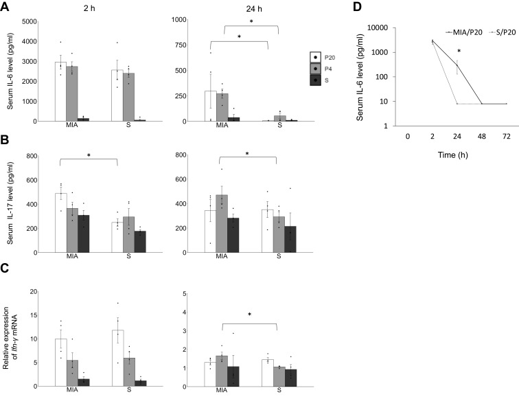
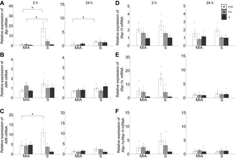
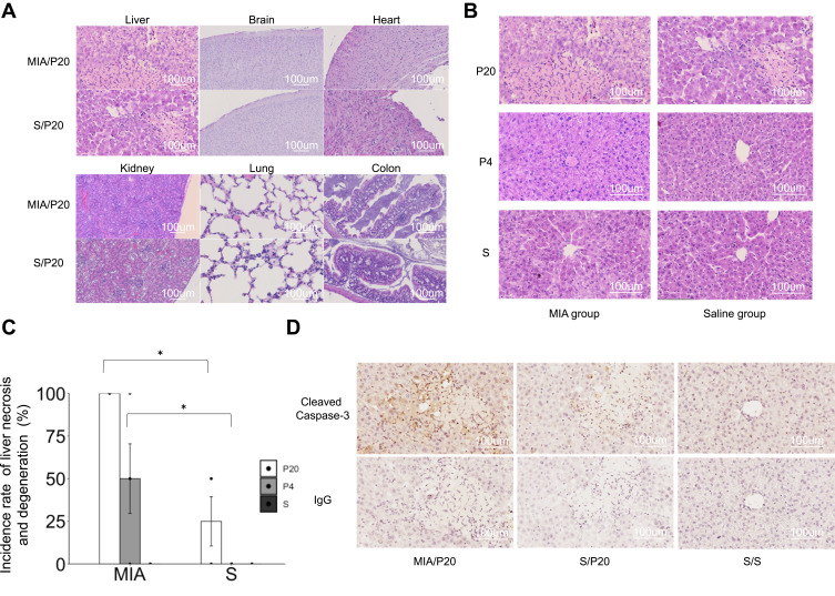
The serum levels of interleukin (IL)-6, IL-17, and interferon-γ were significantly higher in the MIA group, and acute liver necrosis was detected. Moreover, failure in UPR was observed in the MIA group compared with that in the control group.
Overall, MIA exposure in utero caused failure in UPR as well as immune overreaction to the second attack of inflammation in offspring. Our results suggested that prenatal exposure to MIA might contribute to the congenital inflammatory constitution after birth.
已确定多种儿童疾病,如严重感染或自身炎症性疾病,其中针对炎症的免疫过度反应可能是潜在机制。先前的报告表明,暴露于由聚肌苷酸 - 聚胞苷酸[聚(I:C)]诱导的母体免疫激活(MIA)的胎儿细胞在体外对炎症表现出超敏反应。然而,该机制的细节仍不清楚。因此,本研究旨在使用病毒感染小鼠模型揭示产前暴露于MIA的后代对炎症的反应及其分子机制。
在交配后12.5、14.5和16.5天的怀孕小鼠腹腔注射20mg/kg体重(BW)的聚(I:C)或生理盐水。3 - 4周龄的后代接受第二次注射20mg/kg BW或4mg/kg BW的聚(I:C)或生理盐水。在产后注射后2、24、48和72小时收集血清和组织。检测后代的细胞因子谱、器官组织病理学和未折叠蛋白反应(UPR)。
MIA组血清白细胞介素(IL)-6、IL-17和干扰素-γ水平显著升高,并检测到急性肝坏死。此外,与对照组相比,MIA组观察到UPR功能障碍。
总体而言,子宫内暴露于MIA导致后代UPR功能障碍以及对第二次炎症攻击的免疫过度反应。我们的结果表明,产前暴露于MIA可能导致出生后先天性炎症体质。