Cepeda Santiago, García-García Sergio, Arrese Ignacio, Fernández-Pérez Gabriel, Velasco-Casares María, Fajardo-Puentes Manuel, Zamora Tomás, Sarabia Rosario
Neurosurgery Department, University Hospital Río Hortega, Valladolid, Spain.
Radiology Department, University Hospital Río Hortega, Valladolid, Spain.
Front Oncol. 2021 Feb 2;10:590756. doi: 10.3389/fonc.2020.590756. eCollection 2020.
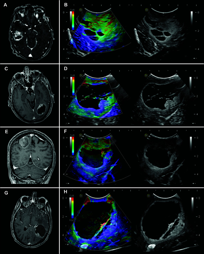
The differential diagnosis of glioblastomas (GBM) from solitary brain metastases (SBM) is essential because the surgical strategy varies according to the histopathological diagnosis. Intraoperative ultrasound elastography (IOUS-E) is a relatively novel technique implemented in the surgical management of brain tumors that provides additional information about the elasticity of tissues. This study compares the discriminative capacity of intraoperative ultrasound B-mode and strain elastography to differentiate GBM from SBM.
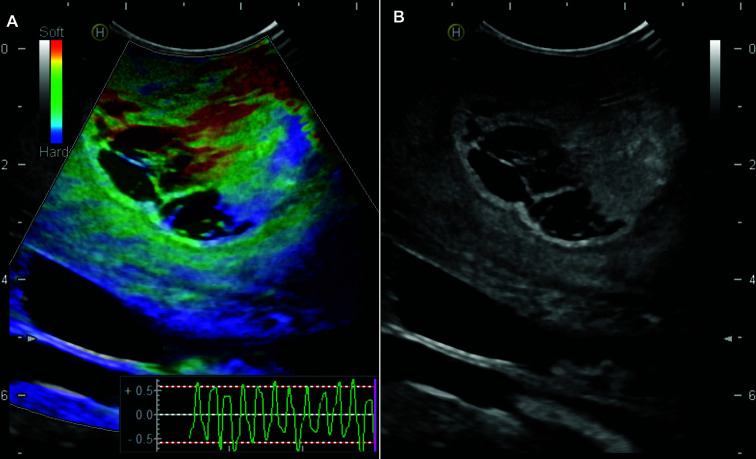
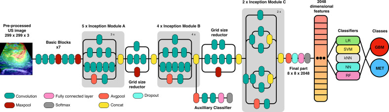
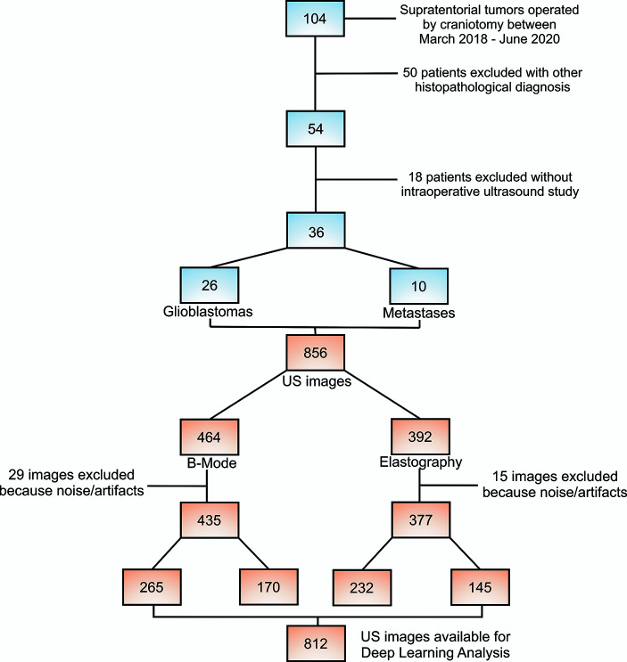
We performed a retrospective analysis of patients who underwent craniotomy between March 2018 to June 2020 with glioblastoma (GBM) and solitary brain metastases (SBM) diagnoses. Cases with an intraoperative ultrasound study were included. Images were acquired before dural opening, first in B-mode, and then using the strain elastography module. After image pre-processing, an analysis based on deep learning was conducted using the open-source software Orange. We have trained an existing neural network to classify tumors into GBM and SBM the transfer learning method using Inception V3. Then, logistic regression (LR) with LASSO (least absolute shrinkage and selection operator) regularization, support vector machine (SVM), random forest (RF), neural network (NN), and k-nearest neighbor (kNN) were used as classification algorithms. After the models' training, ten-fold stratified cross-validation was performed. The models were evaluated using the area under the curve (AUC), classification accuracy, and precision.
A total of 36 patients were included in the analysis, 26 GBM and 10 SBM. Models were built using a total of 812 ultrasound images, 435 of B-mode, 265 (60.92%) corresponded to GBM and 170 (39.8%) to metastases. In addition, 377 elastograms, 232 (61.54%) GBM and 145 (38.46%) metastases were analyzed. For B-mode, AUC and accuracy values of the classification algorithms ranged from 0.790 to 0.943 and from 72 to 89%, respectively. For elastography, AUC and accuracy values ranged from 0.847 to 0.985 and from 79% to 95%, respectively.
Automated processing of ultrasound images through deep learning can generate high-precision classification algorithms that differentiate glioblastomas from metastases using intraoperative ultrasound. The best performance regarding AUC was achieved by the elastography-based model supporting the additional diagnostic value that this technique provides.
胶质母细胞瘤(GBM)与孤立性脑转移瘤(SBM)的鉴别诊断至关重要,因为手术策略会根据组织病理学诊断而有所不同。术中超声弹性成像(IOUS-E)是一种在脑肿瘤手术管理中应用的相对新颖的技术,可提供有关组织弹性的额外信息。本研究比较了术中超声B模式和应变弹性成像区分GBM与SBM的鉴别能力。
我们对2018年3月至2020年6月期间接受开颅手术且诊断为胶质母细胞瘤(GBM)和孤立性脑转移瘤(SBM)的患者进行了回顾性分析。纳入有术中超声检查的病例。在硬脑膜打开前采集图像,首先是B模式,然后使用应变弹性成像模块。图像预处理后,使用开源软件Orange进行基于深度学习的分析。我们训练了一个现有的神经网络,使用迁移学习方法Inception V3将肿瘤分类为GBM和SBM。然后,将带LASSO(最小绝对收缩和选择算子)正则化的逻辑回归(LR)、支持向量机(SVM)、随机森林(RF)、神经网络(NN)和k近邻(kNN)用作分类算法。模型训练后,进行十折分层交叉验证。使用曲线下面积(AUC)、分类准确率和精确率对模型进行评估。
共有36例患者纳入分析,26例GBM和10例SBM。共使用812张超声图像建立模型,其中435张B模式图像,265张(60.92%)对应GBM,170张(39.8%)对应转移瘤。此外,分析了377张弹性成像图,232张(61.54%)GBM和145张(38.46%)转移瘤。对于B模式,分类算法的AUC和准确率值分别为0.790至0.943和72%至89%。对于弹性成像,AUC和准确率值分别为0.847至0.985和79%至95%。
通过深度学习对超声图像进行自动处理,可以生成高精度的分类算法,利用术中超声区分胶质母细胞瘤和转移瘤。基于弹性成像的模型在AUC方面表现最佳,支持该技术提供的额外诊断价值。