Section of Protein Structure and Function, Laboratory of Retinal Cell and Molecular Biology, National Eye Institute, National Institutes of Health, Bethesda, Maryland, United States.
Department of Biochemistry and Molecular & Cellular Biology, Georgetown University Medical Center, Washington DC, United States.
Invest Ophthalmol Vis Sci. 2021 Feb 1;62(2):30. doi: 10.1167/iovs.62.2.30.
To examine the contribution of pigment epithelium-derived factor receptor (PEDF-R) to the phagocytosis process. Previously, we identified PEDF-R, the protein encoded by the PNPLA2 gene, as a phospholipase A2 in the retinal pigment epithelium (RPE). During phagocytosis, RPE cells ingest abundant phospholipids and protein in the form of photoreceptor outer segment (POS) tips, which are then hydrolyzed. The role of PEDF-R in RPE phagocytosis is not known.
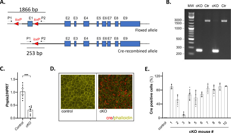
Mice in which PNPLA2 was conditionally knocked out (cKO) in the RPE were generated. Mouse RPE/choroid explants were cultured. Human ARPE-19 cells were transfected with siPNPLA2 silencing duplexes. POSs were isolated from bovine retinas. The phospholipase A2 inhibitor bromoenol lactone was used. Transmission electron microscopy, immunofluorescence, lipid labeling, pulse-chase experiments, western blots, and free fatty acid and β-hydroxybutyrate assays were performed.
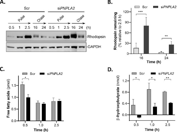
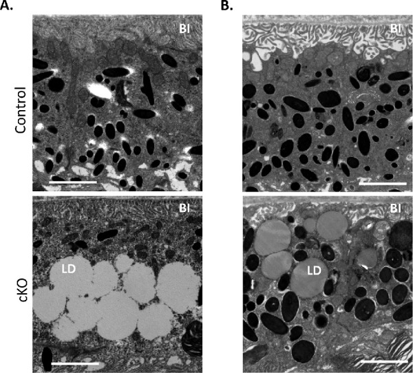
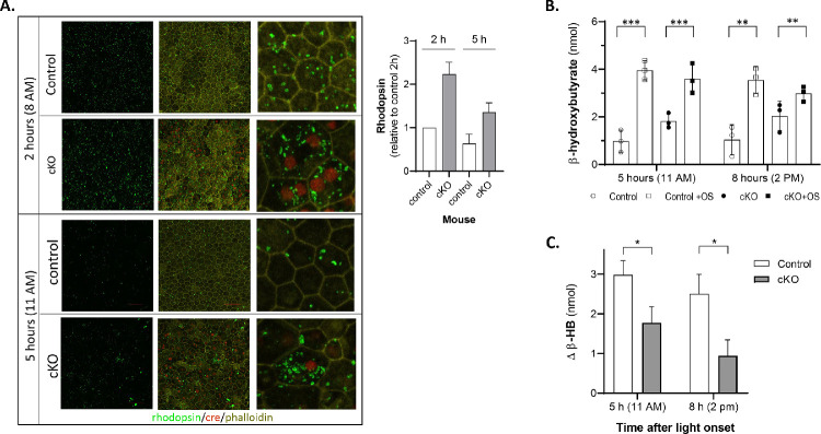
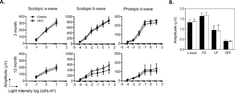
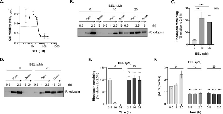
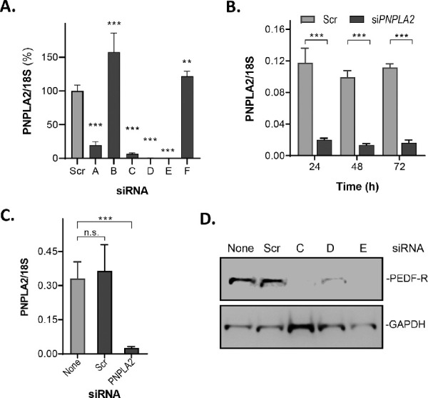
The RPE of the cKO mice accumulated lipids, as well as more abundant and larger rhodopsin particles, compared to littermate controls. Upon POS exposure, RPE explants from cKO mice released less β-hydroxybutyrate compared to controls. After POS ingestion during phagocytosis, rhodopsin degradation was stalled both in cells treated with bromoenol lactone and in PNPLA2-knocked-down cells relative to their corresponding controls. Phospholipase A2 inhibition lowered β-hydroxybutyrate release from phagocytic RPE cells. PNPLA2 knockdown also resulted in a decline in fatty acids and β-hydroxybutyrate release from phagocytic RPE cells.
PEDF-R downregulation delayed POS digestion during phagocytosis. The findings imply that the efficiency of RPE phagocytosis depends on PEDF-R, thus identifying a novel contribution of this protein to POS degradation in the RPE.
研究色素上皮衍生因子受体(PEDF-R)在吞噬过程中的作用。此前,我们鉴定出 PEDF-R 是视网膜色素上皮(RPE)中 PNPLA2 基因编码的蛋白,它是一种磷脂酶 A2。在吞噬过程中,RPE 细胞以光感受器外节(POS)尖端的形式摄取大量磷脂和蛋白质,然后将其水解。PEDF-R 在 RPE 吞噬中的作用尚不清楚。
通过条件性敲除 RPE 中的 PNPLA2(cKO)生成小鼠。培养小鼠 RPE/脉络膜外植体。用 siPNPLA2 沉默寡核苷酸转染人 ARPE-19 细胞。从牛视网膜中分离 POS。使用磷脂酶 A2 抑制剂溴烯内酯。进行透射电子显微镜、免疫荧光、脂质标记、脉冲追踪实验、western blot 以及游离脂肪酸和β-羟丁酸测定。
与同窝对照相比,cKO 小鼠的 RPE 积累了更多的脂质和更大的视紫红质颗粒。与对照相比,cKO 小鼠的 RPE 外植体在暴露于 POS 后释放的β-羟丁酸较少。在吞噬过程中摄入 POS 后,与相应对照相比,用溴烯内酯处理的细胞和敲低 PNPLA2 的细胞中视紫红质降解都停滞。磷脂酶 A2 抑制降低了吞噬性 RPE 细胞中β-羟丁酸的释放。PNPLA2 敲低也导致吞噬性 RPE 细胞中脂肪酸和β-羟丁酸的释放减少。
PEDF-R 下调延迟了吞噬过程中 POS 的消化。这些发现表明,RPE 吞噬的效率取决于 PEDF-R,从而确定了该蛋白对 RPE 中 POS 降解的新贡献。