Zhao Jing, Chen Shuochun, Zhu Lu, Zhang Liang, Liu Jingqi, Xu Danxia, Tian Guo, Jiang Tian'an
Department of Ultrasound, The First Affiliated Hospital, College of Medicine, Zhejiang University, Hangzhou, China.
Key Laboratory of Organ Transplantation, Research Center for Diagnosis and Treatment of Hepatobiliary Diseases, Hangzhou, China.
Front Oncol. 2021 Feb 9;10:621092. doi: 10.3389/fonc.2020.621092. eCollection 2020.
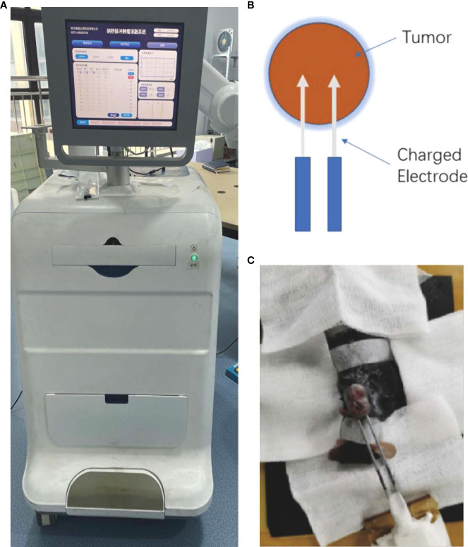
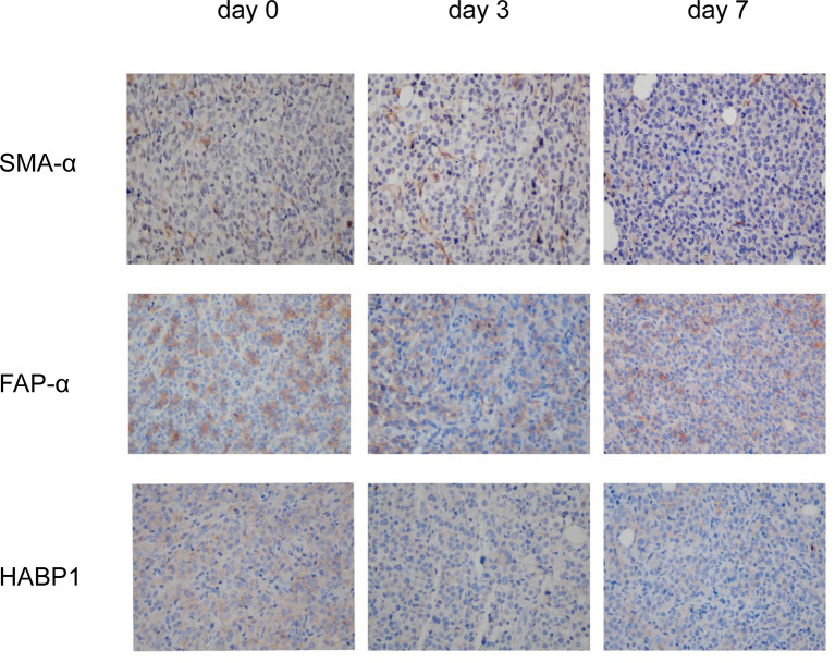
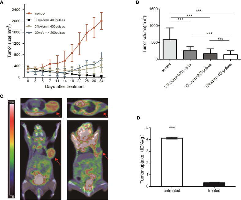
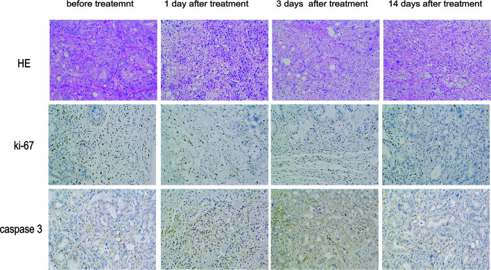
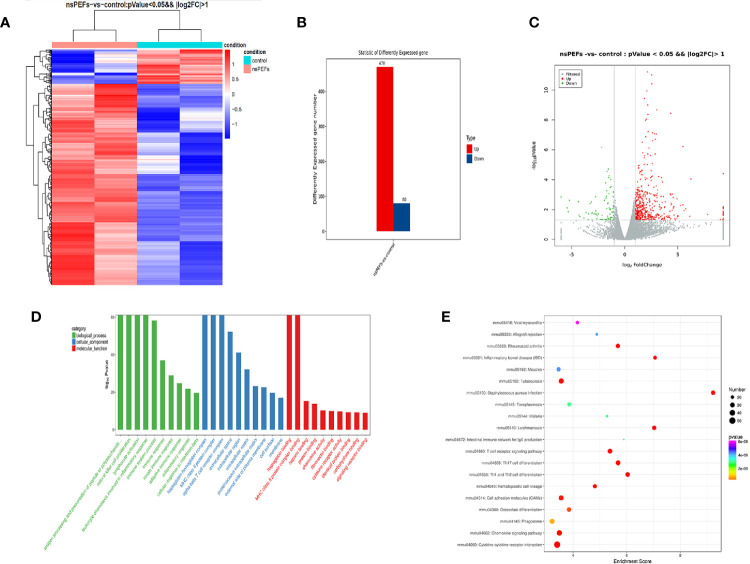
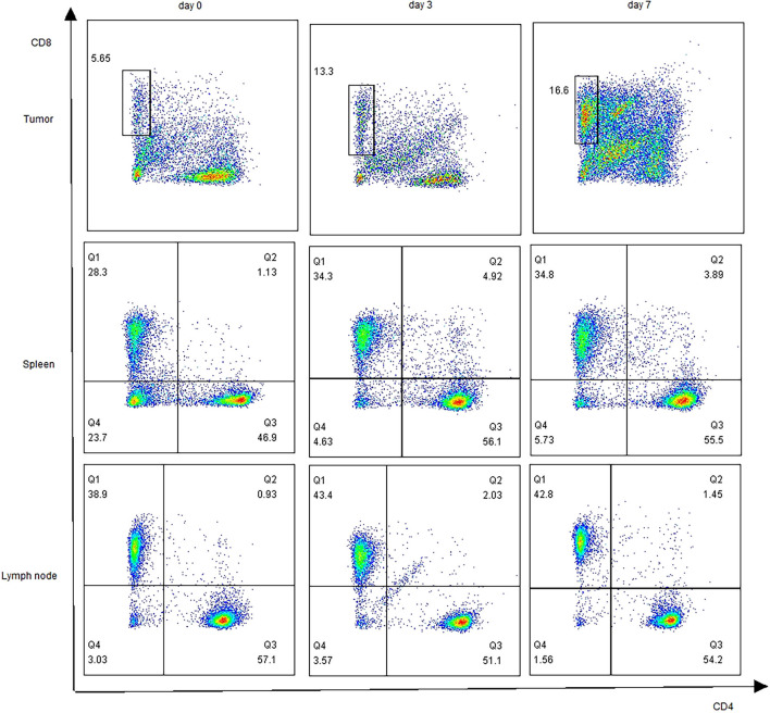
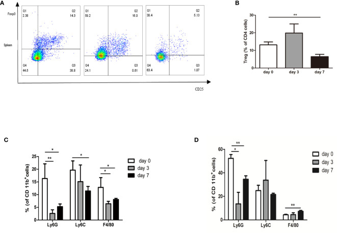
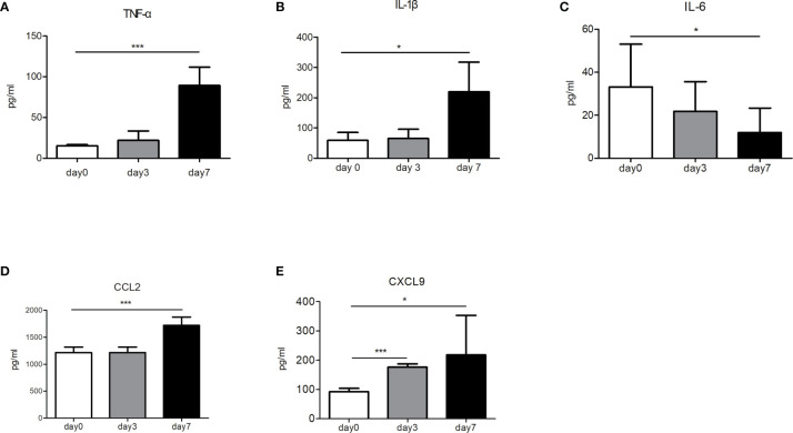
Nanosecond pulsed electric fields (nsPEFs) have emerged as a novel and effective strategy for the non-surgical and minimally invasive removal of tumors. However, the effects of nsPEFs treatment on the tumor immune microenvironment remain unknown. In this study, the changes in the morphology and function of pancreatic cancer cells after nsPEFs were assessed and the modifications in the immune profile in pancreatic cancer models were investigated. To this end, electrodes were inserted with different parameters applied to ablate the targeted tumor tissues. Tumor development was found to be inhibited, with decreased volumes post-nsPEFs treatment compared with control tumors (P < 0.05). Hematoxylin and eosin staining showed morphological changes in pancreatic cancer cells, Ki-67 staining confirmed the effects of nsPEFs on tumor growth, and caspase-3 staining indicated that nsPEFs caused apoptosis in the early stages after treatment. Three days after nsPEFs, positron emission tomography demonstrated little residual metabolic activity compared with the control group. Gene expression profiling identified significant changes in immune-related pathways. After treatment with nsPEFs, CD8 T lymphocytes increased. We showed that nsPEFs led to a significant decrease in immune suppressive cells, including myeloid derived suppressor cells, T regulatory cells, and tumor-associated macrophages. In addition, the levels of TNF-α and IL-1β increased (P < 0.05), while the level of IL-6 was decreased (P < 0.05). NsPEFs alleviated the immunosuppressive components in pancreatic cancer stroma, including hyaluronic acid and fibroblast activation protein-α. Our data demonstrate that tumor growth can be effectively inhibited by nsPEFs . NsPEFs significantly altered the infiltration of immune cells and triggered immune response.
纳秒脉冲电场(nsPEFs)已成为一种用于非手术和微创肿瘤切除的新型有效策略。然而,nsPEFs治疗对肿瘤免疫微环境的影响尚不清楚。在本研究中,评估了nsPEFs作用后胰腺癌细胞的形态和功能变化,并研究了胰腺癌模型中免疫特征的改变。为此,插入电极并应用不同参数来消融靶向肿瘤组织。发现肿瘤生长受到抑制,与对照肿瘤相比,nsPEFs治疗后肿瘤体积减小(P<0.05)。苏木精和伊红染色显示胰腺癌细胞的形态变化,Ki-67染色证实了nsPEFs对肿瘤生长的影响,caspase-3染色表明nsPEFs在治疗后早期引起细胞凋亡。nsPEFs治疗三天后,正电子发射断层扫描显示与对照组相比残留代谢活性较低。基因表达谱分析确定了免疫相关途径的显著变化。nsPEFs治疗后,CD8 T淋巴细胞增加。我们发现nsPEFs导致免疫抑制细胞显著减少,包括骨髓来源的抑制细胞、调节性T细胞和肿瘤相关巨噬细胞。此外,TNF-α和IL-1β水平升高(P<0.05),而IL-6水平降低(P<0.05)。nsPEFs减轻了胰腺癌基质中的免疫抑制成分,包括透明质酸和成纤维细胞活化蛋白-α。我们的数据表明,nsPEFs可以有效抑制肿瘤生长。nsPEFs显著改变了免疫细胞的浸润并引发免疫反应。