Gehrsitz Pia, Rompel Oliver, Schöber Martin, Cesnjevar Robert, Purbojo Ariawan, Uder Michael, Dittrich Sven, Alkassar Muhannad
Department of Pediatric Cardiology, University Hospital Erlangen, Friedrich-Alexander University Erlangen-Nürnberg (FAU), Erlangen, Germany.
Institute of Radiology, University Hospital Erlangen, Friedrich-Alexander University Erlangen-Nürnberg (FAU), Erlangen, Germany.
Front Cardiovasc Med. 2021 Feb 9;8:633611. doi: 10.3389/fcvm.2021.633611. eCollection 2021.
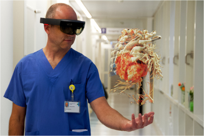
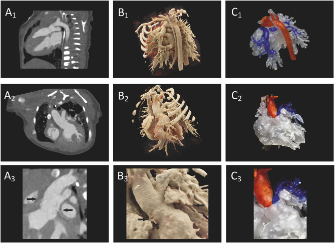
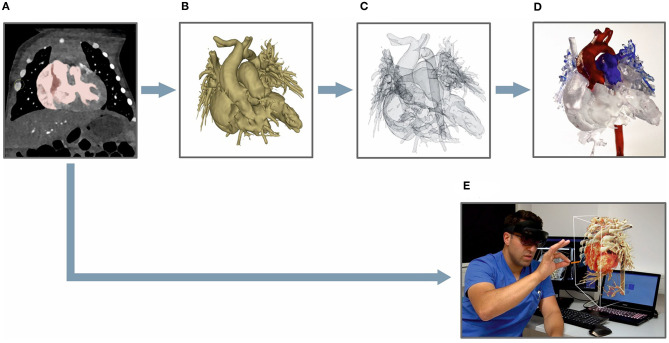
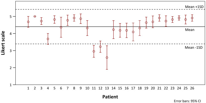
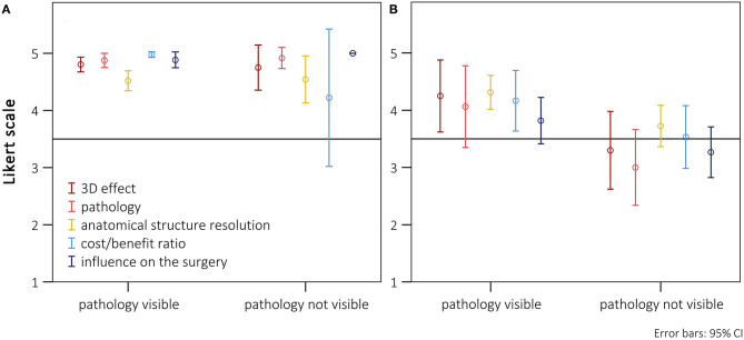
Cinematic rendering (CR) is based on a new algorithm that creates a photo-realistic three-dimensional (3D) picture from cross-sectional images. Previous studies have shown its positive impact on preoperative planning. To date, CR presentation has only been possible on 2D screens which limited natural 3D perception. To depict CR-hearts spatially, we used mixed-reality technology and mapped corresponding hearts as holograms in 3D space. Our aim was to assess the benefits of CR-holograms in the preoperative planning of cardiac surgery. Including 3D prints allowed a direct comparison of two spatially resolved display methods. Twenty-six patients were recruited between February and September 2019. CT or MRI was used to visualize the patient's heart preoperatively. The surgeon was shown the anatomy in cross-sections on a 2D screen, followed by spatial representations as a 3D print and as a high-resolution hologram. The holographic representation was carried out using mixed-reality glasses (HoloLens®). To create the 3D prints, corresponding structures were segmented to create STL files which were printed out of resin. In 22 questions, divided in 5 categories (3D-imaging effect, representation of pathology, structure resolution, cost/benefit ratio, influence on surgery), the surgeons compared each spatial representation with the 2D method, using a five-level Likert scale. The surgical preparation time was assessed by comparing retrospectively matched patient pairs, using a paired -test. CR-holograms surpassed 2D-monitor imaging in all categories. CR-holograms were superior to 3D prints in all categories (mean Likert scale 4.4 ± 1.0 vs. 3.7 ± 1.3, < 0.05). Compared to 3D prints it especially improved the depth perception (4.7 ± 0.7 vs. 3.7 ± 1.2) and the representation of the pathology (4.4 ± 0.9 vs. 3.6 ± 1.2). 3D imaging reduced the intraoperative preparation time ( = 24, 59 ± 23 min vs. 73 ± 43 min, < 0.05). In conclusion, the combination of an extremely photo-realistic presentation via cinematic rendering and the spatial presentation in 3D space via mixed-reality technology allows a previously unattained level of comprehension of anatomy and pathology in preoperative planning.
电影渲染(CR)基于一种新算法,该算法可从横截面图像创建逼真的三维(3D)图片。先前的研究已表明其对术前规划有积极影响。迄今为止,CR呈现仅能在二维屏幕上进行,这限制了自然的3D感知。为了在空间上描绘CR心脏,我们使用了混合现实技术,并将相应的心脏映射为3D空间中的全息图。我们的目的是评估CR全息图在心脏手术术前规划中的益处。纳入3D打印可对两种空间分辨显示方法进行直接比较。2019年2月至9月招募了26名患者。术前使用CT或MRI对患者心脏进行可视化。在二维屏幕上向外科医生展示横截面的解剖结构,随后以3D打印和高分辨率全息图的形式进行空间呈现。使用混合现实眼镜(HoloLens®)进行全息呈现。为了制作3D打印件,对相应结构进行分割以创建STL文件,然后用树脂打印出来。在22个问题中,分为5个类别(3D成像效果、病理表现、结构分辨率、成本效益比、对手术的影响),外科医生使用五级李克特量表将每种空间呈现与二维方法进行比较。通过比较回顾性匹配的患者对来评估手术准备时间,使用配对t检验。CR全息图在所有类别中均超过二维显示器成像。CR全息图在所有类别中均优于3D打印(平均李克特量表4.4±1.0对3.7±1.3,P<0.05)。与3D打印相比,它尤其改善了深度感知(4.7±0.7对3.7±1.2)和病理表现(4.4±0.9对3.6±1.2)。3D成像减少了术中准备时间(n = 24,59±23分钟对73±43分钟,P<0.05)。总之,通过电影渲染实现的极其逼真的呈现与通过混合现实技术在3D空间中的空间呈现相结合,在术前规划中实现了前所未有的解剖结构和病理理解水平。