Fortuni Federico, Hirasawa Kensuke, Bax Jeroen J, Delgado Victoria, Ajmone Marsan Nina
Department of Cardiology, Leiden University Medical Center, Leiden, Netherlands.
Front Cardiovasc Med. 2021 Feb 9;8:638487. doi: 10.3389/fcvm.2021.638487. eCollection 2021.
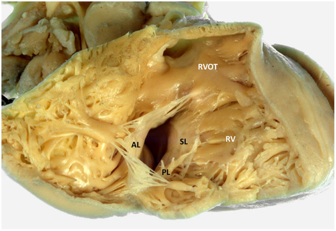
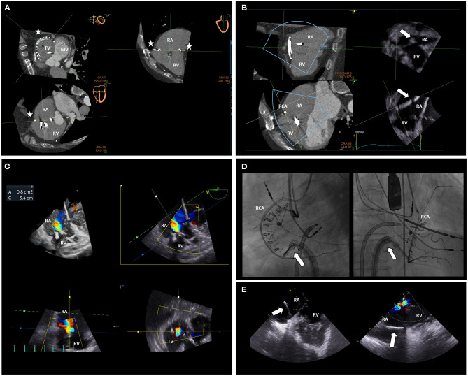
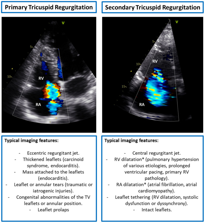
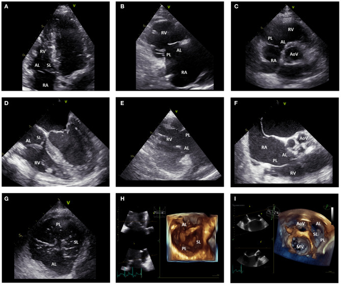
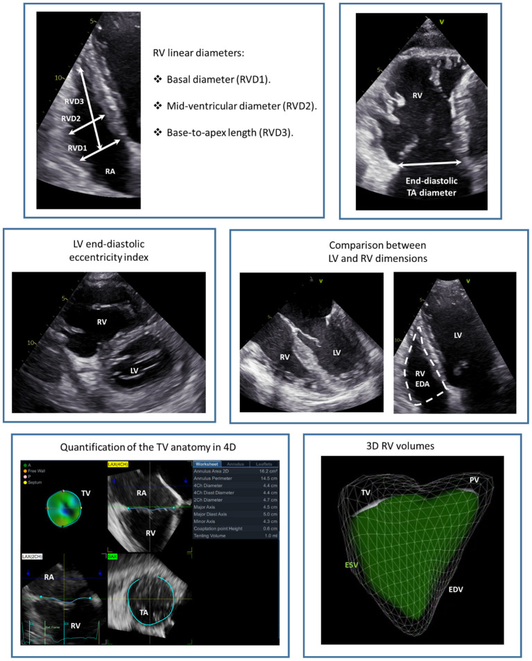
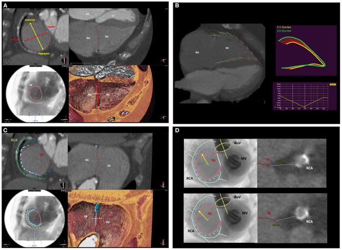
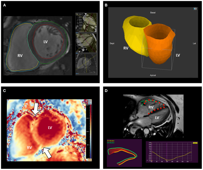
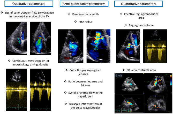
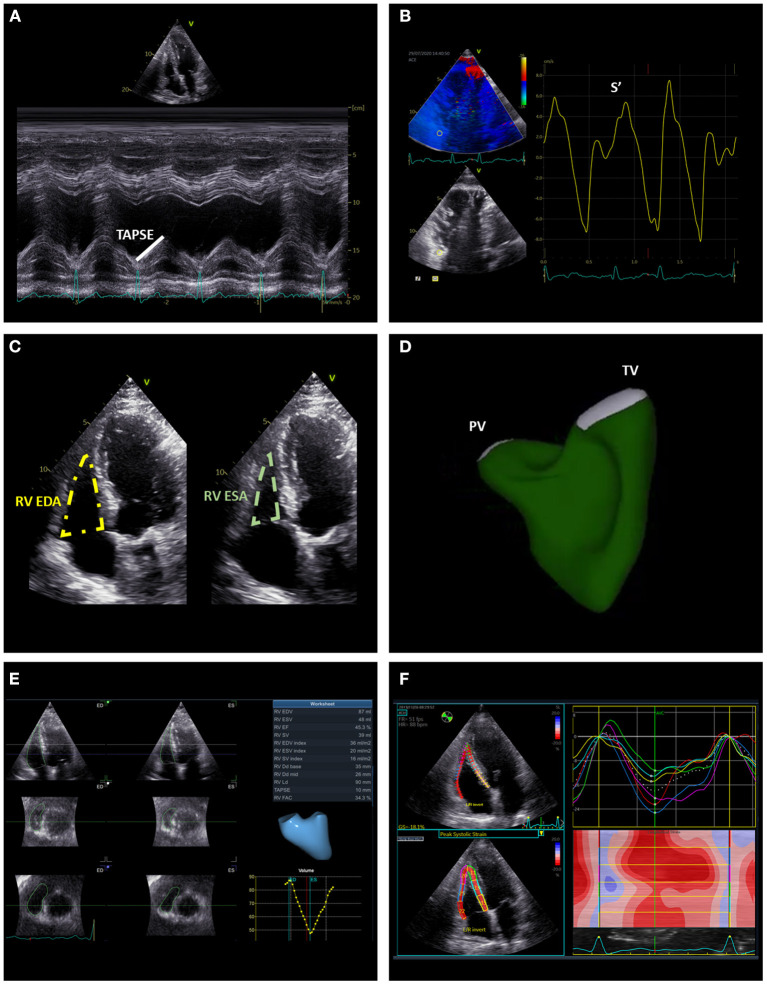
Several studies have demonstrated that severe tricuspid regurgitation (TR) has a significant negative impact on morbidity and mortality. Nowadays, several therapeutic options to treat TR are available and patients at high surgical risk can also be treated with transcatheter procedures. For the management of patients with TR, an accurate assessment of the tricuspid valve and its surrounding structures is therefore of crucial importance and has gained significant interest in the medical community. Different imaging modalities can provide detailed information on the tricuspid valve apparatus, right ventricle, right atrium, and coronary circulation which are fundamental to define the timing and anatomic suitability of surgical and percutaneous procedures. The present review illustrates the role of 2D and 3D echocardiography, cardiac magnetic resonance, and multidetector row computed tomography for the assessment of the tricuspid valve and right heart with a particular focus on the data needed for planning and guiding interventional procedures.
多项研究表明,严重三尖瓣反流(TR)对发病率和死亡率有显著负面影响。如今,有多种治疗TR的方法可供选择,手术风险高的患者也可通过经导管手术进行治疗。因此,对于TR患者的管理,准确评估三尖瓣及其周围结构至关重要,并已引起医学界的广泛关注。不同的成像方式可以提供有关三尖瓣装置、右心室、右心房和冠状动脉循环的详细信息,这些信息对于确定手术和经皮手术的时机及解剖学适宜性至关重要。本综述阐述了二维和三维超声心动图、心脏磁共振成像以及多排螺旋计算机断层扫描在评估三尖瓣和右心方面的作用,特别关注规划和指导介入手术所需的数据。