• 文献检索
  • 文档翻译
  • 深度研究
  • 学术资讯
  • Suppr Zotero 插件Zotero 插件
  • 邀请有礼
  • 套餐&价格
  • 历史记录
应用&插件
Suppr Zotero 插件Zotero 插件浏览器插件Mac 客户端Windows 客户端微信小程序
定价
高级版会员购买积分包购买API积分包
服务
文献检索文档翻译深度研究API 文档MCP 服务
关于我们
关于 Suppr公司介绍联系我们用户协议隐私条款
关注我们

Suppr 超能文献

核心技术专利:CN118964589B侵权必究
粤ICP备2023148730 号-1Suppr @ 2026

文献检索

告别复杂PubMed语法,用中文像聊天一样搜索,搜遍4000万医学文献。AI智能推荐,让科研检索更轻松。

立即免费搜索

文件翻译

保留排版,准确专业,支持PDF/Word/PPT等文件格式,支持 12+语言互译。

免费翻译文档

深度研究

AI帮你快速写综述,25分钟生成高质量综述,智能提取关键信息,辅助科研写作。

立即免费体验

用于角膜内皮细胞培养和组织工程的生物材料。

Biomaterials for corneal endothelial cell culture and tissue engineering.

作者信息

Parekh Mohit, Romano Vito, Hassanin Kareem, Testa Valeria, Wongvisavavit Rintra, Ferrari Stefano, Haneef Atikah, Willoughby Colin, Ponzin Diego, Jhanji Vishal, Sharma Namrata, Daniels Julie, Kaye Stephen B, Ahmad Sajjad, Levis Hannah J

机构信息

Faculty of Brain Sciences, Institute of Ophthalmology, University College London, London, UK.

International Center for Ocular Physiopathology, Fondazione Banca degli Occhi del Veneto Onlus, Venice, Italy.

出版信息

J Tissue Eng. 2021 Feb 16;12:2041731421990536. doi: 10.1177/2041731421990536. eCollection 2021 Jan-Dec.

DOI:10.1177/2041731421990536
PMID:33643603
原文链接:https://pmc.ncbi.nlm.nih.gov/articles/PMC7894589/
Abstract

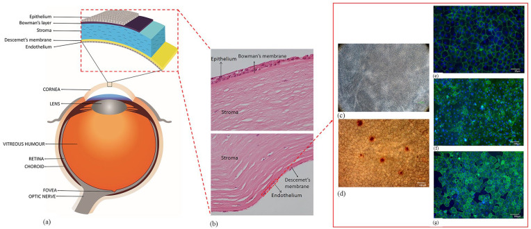
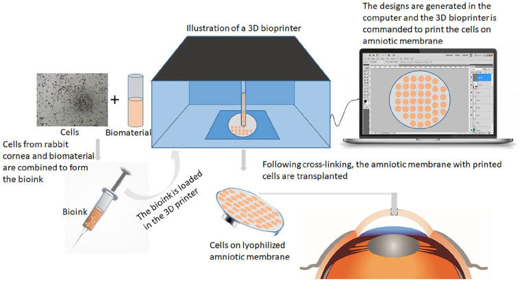
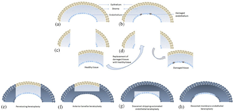
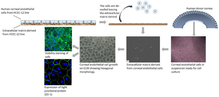
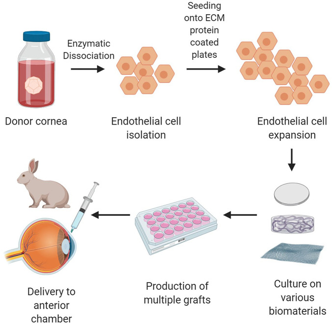
The corneal endothelium is the posterior monolayer of cells that are responsible for maintaining overall transparency of the avascular corneal tissue via pump function. These cells are non-regenerative in vivo and therefore, approximately 40% of corneal transplants undertaken worldwide are a result of damage or dysfunction of endothelial cells. The number of available corneal donor tissues is limited worldwide, hence, cultivation of human corneal endothelial cells (hCECs) in vitro has been attempted in order to produce tissue engineered corneal endothelial grafts. Researchers have attempted to recreate the current gold standard treatment of replacing the endothelial layer with accompanying Descemet's membrane or a small portion of stroma as support with tissue engineering strategies using various substrates of both biologically derived and synthetic origin. Here we review the potential biomaterials that are currently in development to support the transplantation of a cultured monolayer of hCECs.

摘要

角膜内皮是细胞的后单层,其通过泵功能负责维持无血管角膜组织的整体透明度。这些细胞在体内不可再生,因此,全球约40%的角膜移植是内皮细胞损伤或功能障碍的结果。全球可用角膜供体组织数量有限,因此,人们尝试在体外培养人角膜内皮细胞(hCEC),以生产组织工程角膜内皮移植物。研究人员试图用生物衍生和合成来源的各种基质,通过组织工程策略,重现目前用伴随的Descemet膜或一小部分基质作为支撑来替换内皮层的金标准治疗方法。在此,我们综述了目前正在开发的、用于支持培养的单层hCEC移植的潜在生物材料。

https://cdn.ncbi.nlm.nih.gov/pmc/blobs/ed7d/7894589/992677d8ac3a/10.1177_2041731421990536-fig11.jpg
https://cdn.ncbi.nlm.nih.gov/pmc/blobs/ed7d/7894589/88c5b1ce52fe/10.1177_2041731421990536-fig1.jpg
https://cdn.ncbi.nlm.nih.gov/pmc/blobs/ed7d/7894589/6c1554356a93/10.1177_2041731421990536-fig2.jpg
https://cdn.ncbi.nlm.nih.gov/pmc/blobs/ed7d/7894589/37b843c5b8ff/10.1177_2041731421990536-fig3.jpg
https://cdn.ncbi.nlm.nih.gov/pmc/blobs/ed7d/7894589/1474392ddb2a/10.1177_2041731421990536-fig4.jpg
https://cdn.ncbi.nlm.nih.gov/pmc/blobs/ed7d/7894589/a19a8a68e8dc/10.1177_2041731421990536-fig5.jpg
https://cdn.ncbi.nlm.nih.gov/pmc/blobs/ed7d/7894589/6c81c203d50a/10.1177_2041731421990536-fig6.jpg
https://cdn.ncbi.nlm.nih.gov/pmc/blobs/ed7d/7894589/ba882bc709f7/10.1177_2041731421990536-fig7.jpg
https://cdn.ncbi.nlm.nih.gov/pmc/blobs/ed7d/7894589/7b35db8ffdf0/10.1177_2041731421990536-fig8.jpg
https://cdn.ncbi.nlm.nih.gov/pmc/blobs/ed7d/7894589/e4cf3b230b0e/10.1177_2041731421990536-fig9.jpg
https://cdn.ncbi.nlm.nih.gov/pmc/blobs/ed7d/7894589/0edddc58d6c3/10.1177_2041731421990536-fig10.jpg
https://cdn.ncbi.nlm.nih.gov/pmc/blobs/ed7d/7894589/992677d8ac3a/10.1177_2041731421990536-fig11.jpg

相似文献

1
Biomaterials for corneal endothelial cell culture and tissue engineering.用于角膜内皮细胞培养和组织工程的生物材料。
J Tissue Eng. 2021 Feb 16;12:2041731421990536. doi: 10.1177/2041731421990536. eCollection 2021 Jan-Dec.
2
[Transplantation of corneal endothelial cells].[角膜内皮细胞移植]
Nippon Ganka Gakkai Zasshi. 2002 Dec;106(12):805-35; discussion 836.
3
Use of biomaterials in corneal endothelial repair.生物材料在角膜内皮修复中的应用。
Ther Adv Ophthalmol. 2021 Dec 23;13:25158414211058249. doi: 10.1177/25158414211058249. eCollection 2021 Jan-Dec.
4
Tissue engineering of corneal endothelium.角膜内皮的组织工程
J Funct Biomater. 2012 Oct 17;3(4):726-44. doi: 10.3390/jfb3040726.
5
Evaluation of reconstructed human corneal endothelium sheets made with porcine Descemet's membrane in vitro and in vivo.体外和体内评估使用猪 Descemet 膜制作的人角膜内皮细胞片。
Exp Eye Res. 2020 Aug;197:108125. doi: 10.1016/j.exer.2020.108125. Epub 2020 Jul 1.
6
Corneal endothelial regeneration and tissue engineering.角膜内皮细胞的再生与组织工程。
Prog Retin Eye Res. 2013 Jul;35:1-17. doi: 10.1016/j.preteyeres.2013.01.003. Epub 2013 Jan 23.
7
Transplantation of adult human corneal endothelium ex vivo: a morphologic study.成人人类角膜内皮细胞体外移植:一项形态学研究。
Cornea. 2001 Oct;20(7):731-7. doi: 10.1097/00003226-200110000-00012.
8
The impact of biomechanics on corneal endothelium tissue engineering.生物力学对角膜内皮组织工程的影响。
Exp Eye Res. 2021 Aug;209:108690. doi: 10.1016/j.exer.2021.108690. Epub 2021 Jul 1.
9
Decellularized bovine corneal posterior lamellae as carrier matrix for cultivated human corneal endothelial cells.去细胞化牛眼角膜后板作为培养的人眼角膜内皮细胞的载体基质。
Curr Eye Res. 2012 Mar;37(3):179-86. doi: 10.3109/02713683.2011.644382.
10
Micro- and nano-topography to enhance proliferation and sustain functional markers of donor-derived primary human corneal endothelial cells.微米和纳米形貌可增强供体来源的原代人角膜内皮细胞的增殖并维持其功能标记物。
Acta Biomater. 2015 Jun;19:138-48. doi: 10.1016/j.actbio.2015.03.016. Epub 2015 Mar 18.

引用本文的文献

1
Investigating the role of molecular coating in human corneal endothelial cell primary culture using artificial intelligence-driven image analysis.利用人工智能驱动的图像分析研究分子涂层在人角膜内皮细胞原代培养中的作用。
Sci Rep. 2025 Aug 25;15(1):31301. doi: 10.1038/s41598-025-14367-4.
2
Injectable carboxymethyl chitosan/oxidized dextran hydrogels containing zoledronic acid modified strontium hydroxyapatite nanoparticles.含有唑来膦酸修饰的羟基磷灰石纳米颗粒的可注射羧甲基壳聚糖/氧化葡聚糖水凝胶
RSC Adv. 2025 Feb 6;15(6):4014-4028. doi: 10.1039/d4ra08123d.
3
Encountering the clinical complexity of type II Peters anomaly management approaches: a case report.

本文引用的文献

1
Human corneal endothelial cells from older donors can be cultured and passaged on cell-derived extracellular matrix.人眼角膜内皮细胞可从老年供体中培养和传代于细胞衍生的细胞外基质上。
Acta Ophthalmol. 2021 Jun;99(4):e512-e522. doi: 10.1111/aos.14614. Epub 2020 Sep 10.
2
Five-Year Follow-up of First 11 Patients Undergoing Injection of Cultured Corneal Endothelial Cells for Corneal Endothelial Failure.首批11例接受培养角膜内皮细胞注射治疗角膜内皮失代偿患者的五年随访
Ophthalmology. 2021 Apr;128(4):504-514. doi: 10.1016/j.ophtha.2020.09.002. Epub 2020 Sep 6.
3
Evolution of therapies for the corneal endothelium: past, present and future approaches.
应对II型彼得斯异常管理方法的临床复杂性:一例病例报告。
Pan Afr Med J. 2024 Oct 22;49:47. doi: 10.11604/pamj.2024.49.47.44754. eCollection 2024.
4
Biomedical Application of MSCs in Corneal Regeneration and Repair.间充质干细胞在角膜再生与修复中的生物医学应用
Int J Mol Sci. 2025 Jan 15;26(2):695. doi: 10.3390/ijms26020695.
5
Preparation and Characterization of Injectable Augmentation Gels Containing Polycaprolactone and Hyaluronic Acid.含聚己内酯和透明质酸的可注射填充凝胶的制备与表征
J Cosmet Dermatol. 2025 Feb;24(2):e16730. doi: 10.1111/jocd.16730. Epub 2024 Dec 16.
6
Advancements in Polymer Biomaterials as Scaffolds for Corneal Endothelium Tissue Engineering.用于角膜内皮组织工程支架的高分子生物材料的进展
Polymers (Basel). 2024 Oct 12;16(20):2882. doi: 10.3390/polym16202882.
7
Replace or Regenerate? Diverse Approaches to Biomaterials for Treating Corneal Lesions.替换还是再生?治疗角膜病变的生物材料的多样方法。
Biomimetics (Basel). 2024 Mar 28;9(4):202. doi: 10.3390/biomimetics9040202.
8
Predicting Long-Term Endothelial Cell Loss after Preloaded Descemet Membrane Endothelial Keratoplasty in Fuchs' Endothelial Corneal Dystrophy: A Mathematical Model.预测富克斯内皮性角膜营养不良患者预充式Descemet膜内皮角膜移植术后的长期内皮细胞丢失:一种数学模型
J Clin Med. 2024 Feb 2;13(3):877. doi: 10.3390/jcm13030877.
9
Temperature Dependence of Membrane Permeability Parameters for Five Cell Types Using Nonideal Thermodynamic Assumptions to Mathematically Model Cryopreservation Protocols.基于非理想热力学假设,利用数学模型模拟冷冻保存方案,计算五种细胞类型的膜渗透参数的温度依赖性。
J Phys Chem B. 2024 Feb 8;128(5):1139-1160. doi: 10.1021/acs.jpcb.3c04534. Epub 2024 Jan 31.
10
Biomaterials used for tissue engineering of barrier-forming cell monolayers in the eye.用于眼部屏障形成细胞单层组织工程的生物材料。
Front Bioeng Biotechnol. 2023 Sep 27;11:1269385. doi: 10.3389/fbioe.2023.1269385. eCollection 2023.
角膜内皮层治疗方法的演进:过去、现在和未来的方法。
Br J Ophthalmol. 2021 Apr;105(4):454-467. doi: 10.1136/bjophthalmol-2020-316149. Epub 2020 Jul 24.
4
Optimisation of a Novel Bio-Substrate as a Treatment for Atrophic Age-Related Macular Degeneration.一种新型生物基质用于治疗萎缩性年龄相关性黄斑变性的优化
Front Bioeng Biotechnol. 2020 May 15;8:456. doi: 10.3389/fbioe.2020.00456. eCollection 2020.
5
Laminin 511 Precoating Promotes the Functional Recovery of Transplanted Corneal Endothelial Cells.层粘连蛋白 511 预涂可促进移植角膜内皮细胞的功能恢复。
Tissue Eng Part A. 2020 Nov;26(21-22):1158-1168. doi: 10.1089/ten.TEA.2020.0047. Epub 2020 Jul 28.
6
Rapid porcine corneal decellularization through the use of sodium N-lauroyl glutamate and supernuclease.通过使用N-月桂酰谷氨酸钠和超核酸酶实现猪角膜的快速去细胞化。
J Tissue Eng. 2019 Sep 11;10:2041731419875876. doi: 10.1177/2041731419875876. eCollection 2019 Jan-Dec.
7
Descemet membrane endothelial keratoplasty (DMEK): an update on safety, efficacy and patient selection.后弹力层内皮角膜移植术(DMEK):安全性、有效性及患者选择的最新进展
Clin Ophthalmol. 2019 Aug 16;13:1549-1557. doi: 10.2147/OPTH.S178473. eCollection 2019.
8
Poly-ε-lysine based hydrogels as synthetic substrates for the expansion of corneal endothelial cells for transplantation.基于聚-ε-赖氨酸的水凝胶作为用于移植的角膜内皮细胞扩增的合成基质。
J Mater Sci Mater Med. 2019 Sep 4;30(9):102. doi: 10.1007/s10856-019-6303-1.
9
REVIEW: Current understanding of the pathogenesis of Fuchs' endothelial corneal dystrophy.综述:对富克斯内皮性角膜营养不良发病机制的当前认识。
Mol Vis. 2019 Jun 5;25:295-310. eCollection 2019.
10
Gelatin Methacrylate (GelMA)-Based Hydrogels for Cell Transplantation: an Effective Strategy for Tissue Engineering.基于甲基丙烯酰化明胶(GelMA)的水凝胶用于细胞移植:组织工程的有效策略。
Stem Cell Rev Rep. 2019 Oct;15(5):664-679. doi: 10.1007/s12015-019-09893-4.