• 文献检索
  • 文档翻译
  • 深度研究
  • 学术资讯
  • Suppr Zotero 插件Zotero 插件
  • 邀请有礼
  • 套餐&价格
  • 历史记录
应用&插件
Suppr Zotero 插件Zotero 插件浏览器插件Mac 客户端Windows 客户端微信小程序
定价
高级版会员购买积分包购买API积分包
服务
文献检索文档翻译深度研究API 文档MCP 服务
关于我们
关于 Suppr公司介绍联系我们用户协议隐私条款
关注我们

Suppr 超能文献

核心技术专利:CN118964589B侵权必究
粤ICP备2023148730 号-1Suppr @ 2026

文献检索

告别复杂PubMed语法,用中文像聊天一样搜索,搜遍4000万医学文献。AI智能推荐,让科研检索更轻松。

立即免费搜索

文件翻译

保留排版,准确专业,支持PDF/Word/PPT等文件格式,支持 12+语言互译。

免费翻译文档

深度研究

AI帮你快速写综述,25分钟生成高质量综述,智能提取关键信息,辅助科研写作。

立即免费体验

基于 CT 图像的自动化放射组学分析预测有症状肾结石自发排出的可能性。

Automated radiomic analysis of CT images to predict likelihood of spontaneous passage of symptomatic renal stones.

机构信息

Department of Radiology, Mayo Clinic, 200 First Street SW, Rochester, MN, 55905, USA.

Department of Laboratory Medicine and Pathology, Division of Nephrology and Hypertension, Mayo Clinic, Rochester, MN, 55905, USA.

出版信息

Emerg Radiol. 2021 Aug;28(4):781-788. doi: 10.1007/s10140-021-01915-4. Epub 2021 Mar 1.

DOI:10.1007/s10140-021-01915-4
PMID:33644833
原文链接:https://pmc.ncbi.nlm.nih.gov/articles/PMC8477419/
Abstract

PURPOSE

To evaluate the ability of a semi-automated radiomic analysis software in predicting the likelihood of spontaneous passage of urinary stones compared with manual measurements.

METHODS

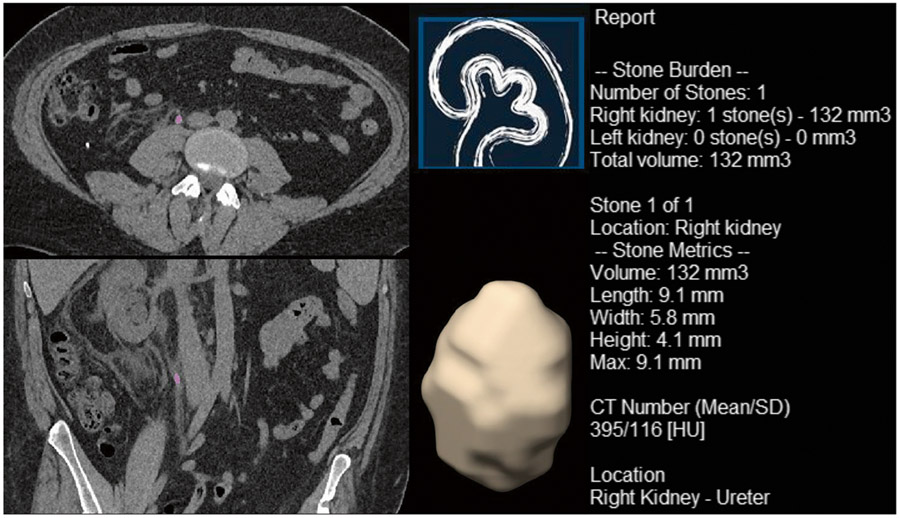
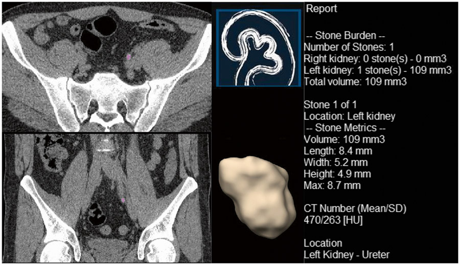
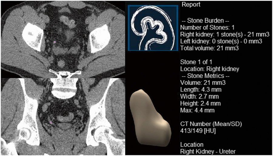
Symptomatic patients visiting the emergency department with suspected stones in either kidney or ureters who underwent a CT scan were included. Patients were followed for up to 6 months for the outcome of a trial of passage. Maximum stone diameters in axial and coronal images were measured manually. Stone length, width, height, max diameter, volume, the mean and standard deviation of the Hounsfield units, and morphologic features were also measured using automated radiomic analysis software. Multivariate models were developed using these data to predict subsequent spontaneous stone passage, with results expressed as the area under a receiver operating curve (AUC).

RESULTS

One hundred eighty-four patients (69 females) with a median age of 56 years were included. Spontaneous stone passage occurred in 114 patients (62%). Univariate analysis demonstrated an AUC of 0.83 and 0.82 for the maximum stone diameter determined manually in the axial and coronal planes, respectively. Multivariate models demonstrated an AUC of 0.82 for a model including manual measurement of maximum stone diameter in axial and coronal planes. The same AUC was found for a model including automatic measurement of maximum height and diameter of the stone. Further addition of morphological parameters measured automatically did not increase AUC beyond 0.83.

CONCLUSION

The semi-automated radiomic analysis of urinary stones shows similar accuracy compared with manual measurements for predicting urinary stone passage. Further studies are needed to predict clinical impacts of reporting the likelihood of urinary stone passage and improving inter-observer variation using automatic radiomic analysis software.

摘要

目的

评估半自动化放射组学分析软件预测尿石自然排出可能性的能力,并与手动测量进行比较。

方法

纳入因疑似肾结石或输尿管结石而就诊于急诊科的症状性患者,所有患者均接受 CT 扫描。对患者进行最长达 6 个月的随访,以确定是否尝试自然排出。手动测量轴向和冠状图像上的最大结石直径。还使用自动化放射组学分析软件测量结石的长度、宽度、高度、最大直径、体积、平均和标准偏差的 Hounsfield 单位以及形态特征。使用这些数据开发多变量模型,以预测随后的自发性结石排出,结果以接受者操作曲线(AUC)下面积表示。

结果

共纳入 184 例(69 例女性)年龄中位数为 56 岁的患者。114 例患者(62%)发生自发性结石排出。单变量分析显示,手动测量轴向和冠状平面最大结石直径的 AUC 分别为 0.83 和 0.82。多变量模型显示,包括轴向和冠状平面最大结石直径手动测量的模型 AUC 为 0.82。包括自动测量结石最大高度和直径的模型也具有相同的 AUC。进一步增加自动测量的形态参数并不能使 AUC 超过 0.83。

结论

与手动测量相比,半自动化放射组学分析在预测尿石排出方面具有相似的准确性。需要进一步的研究来预测使用自动放射组学分析软件报告尿石排出可能性和改善观察者间差异的临床影响。

https://cdn.ncbi.nlm.nih.gov/pmc/blobs/3072/8477419/923c4bb68b59/nihms-1740729-f0003.jpg
https://cdn.ncbi.nlm.nih.gov/pmc/blobs/3072/8477419/aee23b0186ac/nihms-1740729-f0001.jpg
https://cdn.ncbi.nlm.nih.gov/pmc/blobs/3072/8477419/b3cb88d4d99a/nihms-1740729-f0002.jpg
https://cdn.ncbi.nlm.nih.gov/pmc/blobs/3072/8477419/923c4bb68b59/nihms-1740729-f0003.jpg
https://cdn.ncbi.nlm.nih.gov/pmc/blobs/3072/8477419/aee23b0186ac/nihms-1740729-f0001.jpg
https://cdn.ncbi.nlm.nih.gov/pmc/blobs/3072/8477419/b3cb88d4d99a/nihms-1740729-f0002.jpg
https://cdn.ncbi.nlm.nih.gov/pmc/blobs/3072/8477419/923c4bb68b59/nihms-1740729-f0003.jpg

相似文献

1
Automated radiomic analysis of CT images to predict likelihood of spontaneous passage of symptomatic renal stones.基于 CT 图像的自动化放射组学分析预测有症状肾结石自发排出的可能性。
Emerg Radiol. 2021 Aug;28(4):781-788. doi: 10.1007/s10140-021-01915-4. Epub 2021 Mar 1.
2
Prediction of spontaneous ureteral stone passage: Automated 3D-measurements perform equal to radiologists, and linear measurements equal to volumetric.预测自发性输尿管结石排出:自动化 3D 测量与放射科医生相当,线性测量与体积测量相当。
Eur Radiol. 2018 Jun;28(6):2474-2483. doi: 10.1007/s00330-017-5242-9. Epub 2018 Jan 24.
3
CT-based determination of maximum ureteral stone area: a predictor of spontaneous passage.基于 CT 的输尿管结石最大面积测定:预测自然排出的指标。
AJR Am J Roentgenol. 2012 Mar;198(3):603-8. doi: 10.2214/AJR.11.7276.
4
Ureteral Stone Diameter on Computerized Tomography Coronal Reconstructions Is Clinically Important and Under-reported.计算机断层扫描冠状面重建图像上的输尿管结石直径具有临床重要性且报告不足。
Urology. 2017 Apr;102:54-60. doi: 10.1016/j.urology.2016.11.046. Epub 2017 Jan 12.
5
Relationship of spontaneous passage of ureteral calculi to stone size and location as revealed by unenhanced helical CT.非增强螺旋CT显示输尿管结石自然排出与结石大小和位置的关系。
AJR Am J Roentgenol. 2002 Jan;178(1):101-3. doi: 10.2214/ajr.178.1.1780101.
6
Axial Diameter is Superior to Volumetric Measurement in Predicting Ureteral Stone Passage.在预测输尿管结石排出方面,轴径优于容积测量。
Urol Pract. 2021 Sep;8(5):571-575. doi: 10.1097/UPJ.0000000000000242. Epub 2021 Jun 23.
7
Differences in stone size and ureteral dilation between obstructing proximal and distal ureteral calculi.梗阻性近端和远端输尿管结石在结石大小及输尿管扩张方面的差异。
Urology. 2008 Sep;72(3):517-20. doi: 10.1016/j.urology.2008.03.034. Epub 2008 May 27.
8
Predictors for spontaneous stone passage in patients with renal colic secondary to ureteral calculi.预测因输尿管结石引起肾绞痛患者结石自行排出的因素。
Int Urol Nephrol. 2012 Feb;44(1):71-9. doi: 10.1007/s11255-011-9971-4. Epub 2011 May 5.
9
Size matters: The width and location of a ureteral stone accurately predict the chance of spontaneous passage.大小很重要:输尿管结石的宽度和位置准确预测了自行排出的机会。
Eur Radiol. 2017 Nov;27(11):4775-4785. doi: 10.1007/s00330-017-4852-6. Epub 2017 Jun 7.
10
CT-Based Predictors of Spontaneous Ureteral Stone Passage.基于 CT 的输尿管结石自然排出的预测因素。
J Coll Physicians Surg Pak. 2024 Aug;34(8):879-884. doi: 10.29271/jcpsp.2024.08.879.

引用本文的文献

1
Improved display and detection of small renal stones using photon-counting detector CT compared to conventional energy-integrating detector CT.与传统能量积分探测器CT相比,使用光子计数探测器CT可改善小肾结石的显示和检测。
Abdom Radiol (NY). 2025 Jan 27. doi: 10.1007/s00261-024-04781-z.
2
A multicenter study on the application of artificial intelligence radiological characteristics to predict prognosis after percutaneous nephrolithotomy.多中心研究人工智能影像学特征在经皮肾镜取石术后预测预后中的应用。
Front Endocrinol (Lausanne). 2023 Sep 15;14:1184608. doi: 10.3389/fendo.2023.1184608. eCollection 2023.
3
Application of Radiomics Model of CT Images in the Identification of Ureteral Calculus and Phlebolith.

本文引用的文献

1
Morphological characteristics and microstructure of kidney stones using synchrotron radiation μCT reveal the mechanism of crystal growth and aggregation in mixed stones.利用同步辐射微计算机断层扫描对肾结石的形态特征和微观结构进行研究,揭示了混合结石中晶体生长和聚集的机制。
PLoS One. 2019 Mar 22;14(3):e0214003. doi: 10.1371/journal.pone.0214003. eCollection 2019.
2
Prediction of spontaneous ureteral stone passage: Automated 3D-measurements perform equal to radiologists, and linear measurements equal to volumetric.预测自发性输尿管结石排出:自动化 3D 测量与放射科医生相当,线性测量与体积测量相当。
Eur Radiol. 2018 Jun;28(6):2474-2483. doi: 10.1007/s00330-017-5242-9. Epub 2018 Jan 24.
3
CT 图像纹理分析模型在输尿管结石和静脉石鉴别中的应用
Int J Clin Pract. 2022 Nov 14;2022:5478908. doi: 10.1155/2022/5478908. eCollection 2022.
4
Software-Estimated Stone Volume Is Better Predictor of Spontaneous Passage for Acute Nephrolithiasis.软件估算的结石体积是急性肾结石自然排出的更好预测指标。
J Endourol. 2023 Jan;37(1):85-92. doi: 10.1089/end.2022.0475. Epub 2022 Dec 7.
5
Radiomics in Urolithiasis: Systematic Review of Current Applications, Limitations, and Future Directions.尿石症中的放射组学:当前应用、局限性及未来方向的系统评价
J Clin Med. 2022 Aug 31;11(17):5151. doi: 10.3390/jcm11175151.
Size matters: The width and location of a ureteral stone accurately predict the chance of spontaneous passage.
大小很重要:输尿管结石的宽度和位置准确预测了自行排出的机会。
Eur Radiol. 2017 Nov;27(11):4775-4785. doi: 10.1007/s00330-017-4852-6. Epub 2017 Jun 7.
4
Accuracy of ultrasonography for renal stone detection and size determination: is it good enough for management decisions?超声检查对肾结石检测及大小测定的准确性:其对于治疗决策而言是否足够可靠?
BJU Int. 2017 Mar;119(3):464-469. doi: 10.1111/bju.13605. Epub 2016 Aug 17.
5
EAU Guidelines on Diagnosis and Conservative Management of Urolithiasis.EAU 指南:尿石症的诊断和保守治疗管理。
Eur Urol. 2016 Mar;69(3):468-74. doi: 10.1016/j.eururo.2015.07.040. Epub 2015 Aug 28.
6
Analysis of factors affecting spontaneous expulsion of ureteral stones that may predict unfavorable outcomes during watchful waiting periods: What is the influence of diabetes mellitus on the ureter?影响输尿管结石自然排出的因素分析,这些因素可能预示观察等待期的不良结局:糖尿病对输尿管有何影响?
Korean J Urol. 2015 Jun;56(6):455-60. doi: 10.4111/kju.2015.56.6.455. Epub 2015 May 27.
7
Usefulness of hounsfield unit and density in the assessment and treatment of urinary stones.亨氏单位和密度在尿路结石评估与治疗中的应用价值
World J Nephrol. 2014 Nov 6;3(4):282-6. doi: 10.5527/wjn.v3.i4.282.
8
Measurement of ureteric stone diameter in different planes on multidetector computed tomography--impact on the clinical decision making.多排螺旋 CT 不同层面测量输尿管结石直径对临床决策的影响。
Urology. 2014 Feb;83(2):288-92. doi: 10.1016/j.urology.2013.09.037. Epub 2013 Nov 22.
9
Management of renal colic.肾绞痛的管理
BMJ. 2012 Aug 29;345:e5499. doi: 10.1136/bmj.e5499.
10
Kidney stone volume estimation from computerized tomography images using a model based method of correcting for the point spread function.基于模型的方法校正点扩散函数,从计算机断层扫描图像估算肾结石体积。
J Urol. 2012 Sep;188(3):989-95. doi: 10.1016/j.juro.2012.04.098. Epub 2012 Jul 21.