Department of Cancer Systems Imaging, University of Texas MD Anderson Cancer Center, Houston, TX 77030, USA.
Cells. 2021 Feb 26;10(3):499. doi: 10.3390/cells10030499.
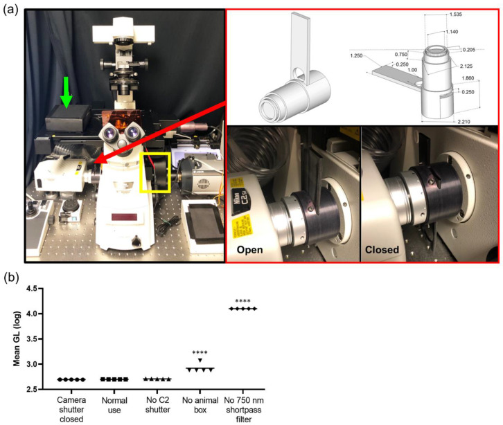
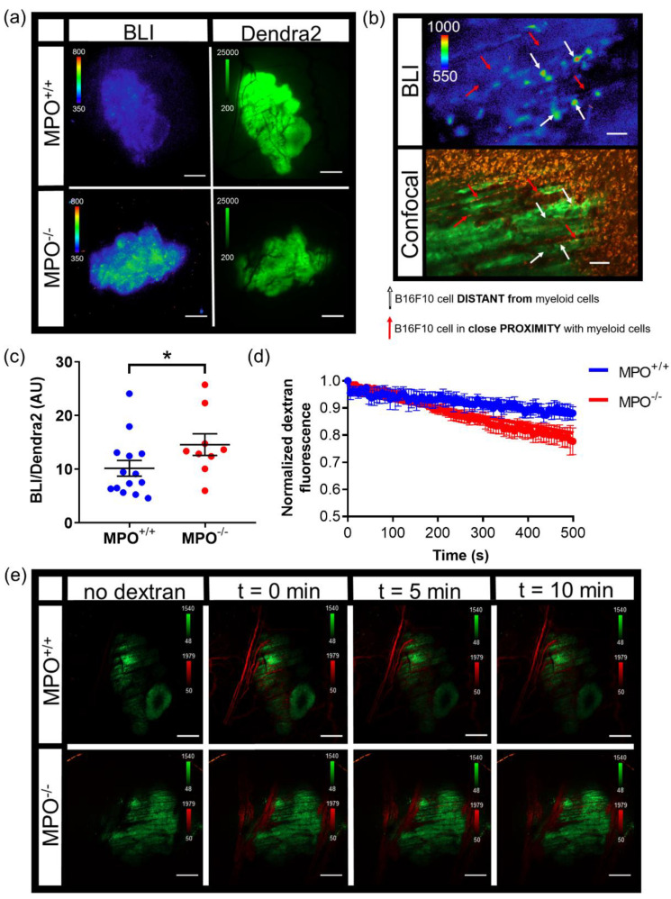
Intravital microscopic imaging (IVM) allows for the study of interactions between immune cells and tumor cells in a dynamic, physiologically relevant system in vivo. Current IVM strategies primarily use fluorescence imaging; however, with the advances in bioluminescence imaging and the development of new bioluminescent reporters with expanded emission spectra, the applications for bioluminescence are extending to single cell imaging. Herein, we describe a molecular imaging window chamber platform that uniquely combines both bioluminescent and fluorescent genetically encoded reporters, as well as exogenous reporters, providing a powerful multi-plex strategy to study molecular and cellular processes in real-time in intact living systems at single cell resolution all in one system. We demonstrate that our molecular imaging window chamber platform is capable of imaging signaling dynamics in real-time at cellular resolution during tumor progression. Importantly, we expand the utility of IVM by modifying an off-the-shelf commercial system with the addition of bioluminescence imaging achieved by the addition of a CCD camera and demonstrate high quality imaging within the reaches of any biology laboratory.
活体显微镜成像(IVM)允许在体内动态、生理相关的系统中研究免疫细胞和肿瘤细胞之间的相互作用。目前的 IVM 策略主要使用荧光成像;然而,随着生物发光成像的进步和具有扩展发射光谱的新型生物发光报告基因的发展,生物发光的应用正在扩展到单细胞成像。在此,我们描述了一种分子成像窗室平台,该平台独特地结合了生物发光和荧光基因编码报告基因,以及外源性报告基因,提供了一种强大的多指标策略,可在单个细胞分辨率下实时研究完整活体系统中的分子和细胞过程。我们证明,我们的分子成像窗室平台能够在肿瘤进展过程中以细胞分辨率实时成像信号动力学。重要的是,我们通过在现成的商业系统中添加生物发光成像来扩展 IVM 的实用性,通过添加 CCD 相机来实现,并且证明了在任何生物学实验室都能达到的高质量成像。