• 文献检索
  • 文档翻译
  • 深度研究
  • 学术资讯
  • Suppr Zotero 插件Zotero 插件
  • 邀请有礼
  • 套餐&价格
  • 历史记录
应用&插件
Suppr Zotero 插件Zotero 插件浏览器插件Mac 客户端Windows 客户端微信小程序
定价
高级版会员购买积分包购买API积分包
服务
文献检索文档翻译深度研究API 文档MCP 服务
关于我们
关于 Suppr公司介绍联系我们用户协议隐私条款
关注我们

Suppr 超能文献

核心技术专利:CN118964589B侵权必究
粤ICP备2023148730 号-1Suppr @ 2026

文献检索

告别复杂PubMed语法,用中文像聊天一样搜索,搜遍4000万医学文献。AI智能推荐,让科研检索更轻松。

立即免费搜索

文件翻译

保留排版,准确专业,支持PDF/Word/PPT等文件格式,支持 12+语言互译。

免费翻译文档

深度研究

AI帮你快速写综述,25分钟生成高质量综述,智能提取关键信息,辅助科研写作。

立即免费体验

相似文献

1
Single-cell meta-analysis of SARS-CoV-2 entry genes across tissues and demographics.单细胞多组学分析 SARS-CoV-2 进入细胞的基因在不同组织和人群中的差异
Nat Med. 2021 Mar;27(3):546-559. doi: 10.1038/s41591-020-01227-z. Epub 2021 Mar 2.
2
Dysbalance of ACE2 levels - a possible cause for severe COVID-19 outcome in COPD.ACE2 水平失衡 - COPD 患者发生严重 COVID-19 结局的一个可能原因。
J Pathol Clin Res. 2021 Sep;7(5):446-458. doi: 10.1002/cjp2.224. Epub 2021 May 12.
3
Contributions of human ACE2 and TMPRSS2 in determining host-pathogen interaction of COVID-19.人类血管紧张素转换酶2(ACE2)和跨膜丝氨酸蛋白酶2(TMPRSS2)在决定新型冠状病毒肺炎(COVID-19)宿主-病原体相互作用中的作用。
J Genet. 2021;100(1). doi: 10.1007/s12041-021-01262-w.
4
Human eggs, zygotes, and embryos express the receptor angiotensin 1-converting enzyme 2 and transmembrane serine protease 2 protein necessary for severe acute respiratory syndrome coronavirus 2 infection.人类卵子、受精卵和胚胎表达血管紧张素转换酶 1 受体和跨膜丝氨酸蛋白酶 2 蛋白,这是严重急性呼吸综合征冠状病毒 2 感染所必需的。
F S Sci. 2021 Feb;2(1):33-42. doi: 10.1016/j.xfss.2020.12.005. Epub 2020 Dec 24.
5
Heterogeneous expression of the SARS-Coronavirus-2 receptor ACE2 in the human respiratory tract.人类呼吸道中 SARS-CoV-2 受体 ACE2 的异质性表达。
EBioMedicine. 2020 Oct;60:102976. doi: 10.1016/j.ebiom.2020.102976. Epub 2020 Sep 21.
6
Expression profiling meta-analysis of ACE2 and TMPRSS2, the putative anti-inflammatory receptor and priming protease of SARS-CoV-2 in human cells, and identification of putative modulators.ACE2 和 TMPRSS2 的表达谱荟萃分析,SARS-CoV-2 在人细胞中的假定抗炎受体和启动蛋白酶,以及假定调节剂的鉴定。
Redox Biol. 2020 Sep;36:101615. doi: 10.1016/j.redox.2020.101615. Epub 2020 Jun 24.
7
SARS-CoV-2 Receptor ACE2 Is an Interferon-Stimulated Gene in Human Airway Epithelial Cells and Is Detected in Specific Cell Subsets across Tissues.SARS-CoV-2 受体 ACE2 是人类气道上皮细胞中的一种干扰素刺激基因,可在组织中的特定细胞亚群中检测到。
Cell. 2020 May 28;181(5):1016-1035.e19. doi: 10.1016/j.cell.2020.04.035. Epub 2020 Apr 27.
8
Gene expression and protein profiling of candidate SARS-CoV-2 receptors in human airway epithelial cells and lung tissue.人类气道上皮细胞和肺组织中候选 SARS-CoV-2 受体的基因表达和蛋白质谱分析。
Eur Respir J. 2020 Sep 3;56(3). doi: 10.1183/13993003.01123-2020. Print 2020 Sep.
9
SARS-CoV-2 pseudovirus infectivity and expression of viral entry-related factors ACE2, TMPRSS2, Kim-1, and NRP-1 in human cells from the respiratory, urinary, digestive, reproductive, and immune systems.SARS-CoV-2 假病毒感染性及其在呼吸道、泌尿道、消化道、生殖系统和免疫系统的人类细胞中病毒进入相关因子 ACE2、TMPRSS2、Kim-1 和 NRP-1 的表达。
J Med Virol. 2021 Dec;93(12):6671-6685. doi: 10.1002/jmv.27244. Epub 2021 Aug 4.
10
and expression by clinical, HLA, immune, and microbial correlates across 34 human cancers and matched normal tissues: implications for SARS-CoV-2 COVID-19.并通过 34 种人类癌症及匹配的正常组织中的临床、HLA、免疫和微生物相关性进行表达:对 SARS-CoV-2 COVID-19 的影响。
J Immunother Cancer. 2020 Jul;8(2). doi: 10.1136/jitc-2020-001020.

引用本文的文献

1
A Benchmark of Semi-Supervised scRNA-seq Integration Methods in Real-World Scenarios.真实场景下半监督单细胞RNA测序整合方法的基准测试
bioRxiv. 2025 Aug 27:2025.08.23.671952. doi: 10.1101/2025.08.23.671952.
2
Addressing Sex as a Biological Variable in Preclinical Models of Lung Disease: An Official American Thoracic Society Research Statement.将性别作为肺部疾病临床前模型中的生物学变量来考量:美国胸科学会官方研究声明
Am J Respir Crit Care Med. 2025 Aug;211(8):1346-1368. doi: 10.1164/rccm.202506-1361ST.
3
Single-cell transcriptome analysis of bronchoalveolar lavage during early SARS-CoV-2 infection.新型冠状病毒2型(SARS-CoV-2)早期感染期间支气管肺泡灌洗的单细胞转录组分析
Microbiol Spectr. 2025 Sep 2;13(9):e0271524. doi: 10.1128/spectrum.02715-24. Epub 2025 Jul 31.
4
Age-dependent ACE2/TMPRSS2 expression and SARS-CoV-2 household transmission in Gran Canaria.大加那利岛年龄依赖性ACE2/TMPRSS2表达与新冠病毒家庭传播情况
PLoS One. 2025 Jul 29;20(7):e0329229. doi: 10.1371/journal.pone.0329229. eCollection 2025.
5
Joint representation and visualization of derailed cell states with Decipher.使用Decipher对脱轨细胞状态进行联合表示和可视化。
Genome Biol. 2025 Jul 23;26(1):219. doi: 10.1186/s13059-025-03682-8.
6
Predicting SARS-CoV-2-specific CD4 and CD8 T-cell responses elicited by inactivated vaccines in healthy adults using machine learning models.使用机器学习模型预测灭活疫苗在健康成年人中引发的SARS-CoV-2特异性CD4和CD8 T细胞反应。
Clin Exp Med. 2025 Jul 8;25(1):236. doi: 10.1007/s10238-025-01772-2.
7
Pathophysiology of COVID-19: A Post Hoc Analysis of the ICAT-COVID Clinical Trial of the Bradykinin Antagonist Icatibant.新型冠状病毒肺炎的病理生理学:缓激肽拮抗剂依卡替班ICAT-COVID临床试验的事后分析
Pathogens. 2025 May 27;14(6):533. doi: 10.3390/pathogens14060533.
8
Infectious and Immunological Links Between Periodontitis and COVID-19: A Review.牙周炎与2019冠状病毒病之间的感染及免疫联系:综述
Med Sci Monit. 2025 May 26;31:e948069. doi: 10.12659/MSM.948069.
9
Determinants of susceptibility to SARS-CoV-2 infection in murine ACE2.小鼠血管紧张素转换酶2(ACE2)对严重急性呼吸综合征冠状病毒2(SARS-CoV-2)感染易感性的决定因素。
J Virol. 2025 Jun 17;99(6):e0054325. doi: 10.1128/jvi.00543-25. Epub 2025 May 12.
10
Human Alveolar Macrophages Detect SARS-CoV-2 Envelope Protein Through TLR2 and TLR4 and Secrete Cytokines in Response.人肺泡巨噬细胞通过TLR2和TLR4检测严重急性呼吸综合征冠状病毒2(SARS-CoV-2)包膜蛋白并分泌细胞因子作为应答。
Immunology. 2025 Jul;175(3):391-401. doi: 10.1111/imm.13922. Epub 2025 May 4.

本文引用的文献

1
A molecular cell atlas of the human lung from single-cell RNA sequencing.人类肺部单细胞 RNA 测序的分子细胞图谱。
Nature. 2020 Nov;587(7835):619-625. doi: 10.1038/s41586-020-2922-4. Epub 2020 Nov 18.
2
An inflammatory cytokine signature predicts COVID-19 severity and survival.炎症细胞因子特征可预测 COVID-19 严重程度和存活情况。
Nat Med. 2020 Oct;26(10):1636-1643. doi: 10.1038/s41591-020-1051-9. Epub 2020 Aug 24.
3
Single-cell RNA-seq reveals ectopic and aberrant lung-resident cell populations in idiopathic pulmonary fibrosis.单细胞 RNA 测序揭示特发性肺纤维化中异位和异常的肺驻留细胞群体。
Sci Adv. 2020 Jul 8;6(28):eaba1983. doi: 10.1126/sciadv.aba1983. eCollection 2020 Jul.
4
Single-cell RNA sequencing reveals profibrotic roles of distinct epithelial and mesenchymal lineages in pulmonary fibrosis.单细胞 RNA 测序揭示了肺纤维化中不同上皮和间充质谱系的促纤维化作用。
Sci Adv. 2020 Jul 8;6(28):eaba1972. doi: 10.1126/sciadv.aba1972. eCollection 2020 Jul.
5
Myocyte-Specific Upregulation of in Cardiovascular Disease: Implications for SARS-CoV-2-Mediated Myocarditis.肌细胞特异性上调在心血管疾病中的作用:对 SARS-CoV-2 介导的心肌炎的影响。
Circulation. 2020 Aug 18;142(7):708-710. doi: 10.1161/CIRCULATIONAHA.120.047911. Epub 2020 Jun 22.
6
A Single-Cell Atlas of the Human Healthy Airways.人类健康气道单细胞图谱
Am J Respir Crit Care Med. 2020 Dec 15;202(12):1636-1645. doi: 10.1164/rccm.201911-2199OC.
7
Serology-informed estimates of SARS-CoV-2 infection fatality risk in Geneva, Switzerland.瑞士日内瓦基于血清学的新型冠状病毒2感染致死风险评估
Lancet Infect Dis. 2021 Apr;21(4):e69-e70. doi: 10.1016/S1473-3099(20)30584-3. Epub 2020 Jul 14.
8
SARS-CoV-2 Reverse Genetics Reveals a Variable Infection Gradient in the Respiratory Tract.SARS-CoV-2 反向遗传学揭示了呼吸道感染的可变梯度。
Cell. 2020 Jul 23;182(2):429-446.e14. doi: 10.1016/j.cell.2020.05.042. Epub 2020 May 27.
9
Knowledge synthesis of 100 million biomedical documents augments the deep expression profiling of coronavirus receptors.对 1 亿篇生物医学文献的知识综合增强了冠状病毒受体的深度表达分析。
Elife. 2020 May 28;9:e58040. doi: 10.7554/eLife.58040.
10
More on Neurologic Features in Severe SARS-CoV-2 Infection. Reply.关于严重SARS-CoV-2感染的神经学特征的更多内容。回复。
N Engl J Med. 2020 Jun 25;382(26):e110. doi: 10.1056/NEJMc2015132. Epub 2020 May 26.

单细胞多组学分析 SARS-CoV-2 进入细胞的基因在不同组织和人群中的差异

Single-cell meta-analysis of SARS-CoV-2 entry genes across tissues and demographics.

机构信息

Klarman Cell Observatory, Broad Institute of MIT and Harvard, Cambridge, MA, USA.

John A. Paulson School of Engineering and Applied Sciences, Harvard University, Cambridge, MA, USA.

出版信息

Nat Med. 2021 Mar;27(3):546-559. doi: 10.1038/s41591-020-01227-z. Epub 2021 Mar 2.

DOI:10.1038/s41591-020-01227-z
PMID:33654293
原文链接:https://pmc.ncbi.nlm.nih.gov/articles/PMC9469728/
Abstract

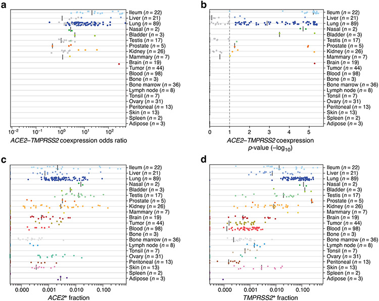
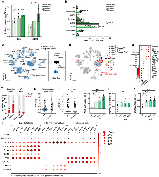
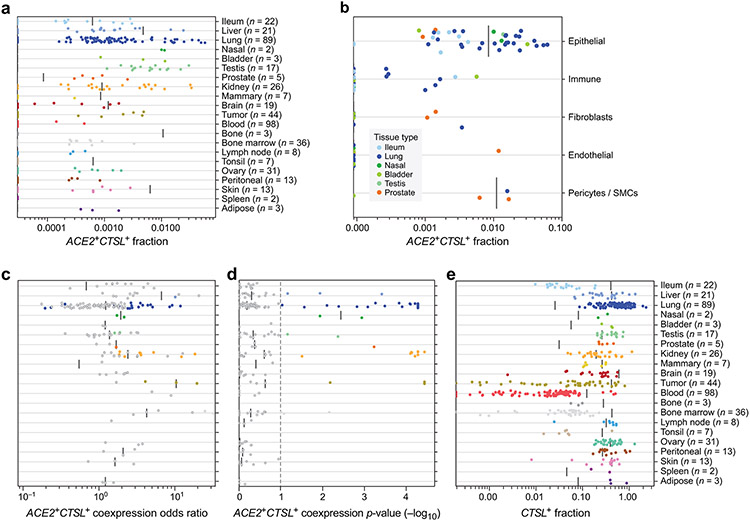
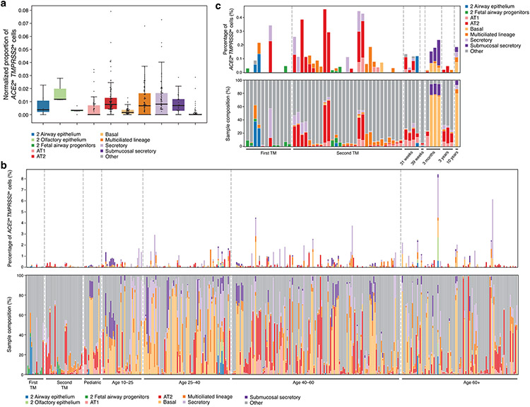
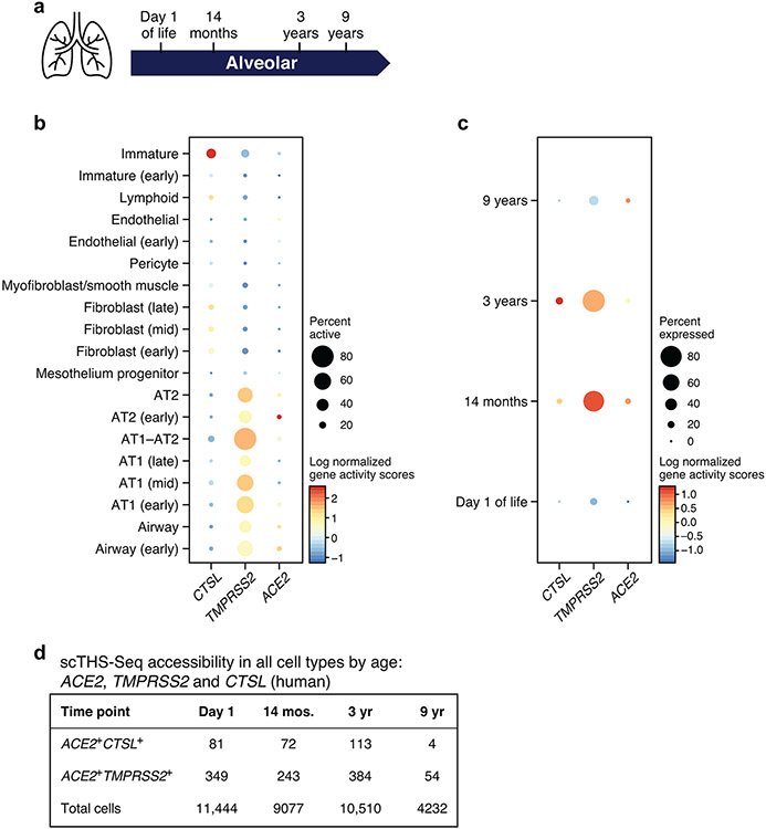
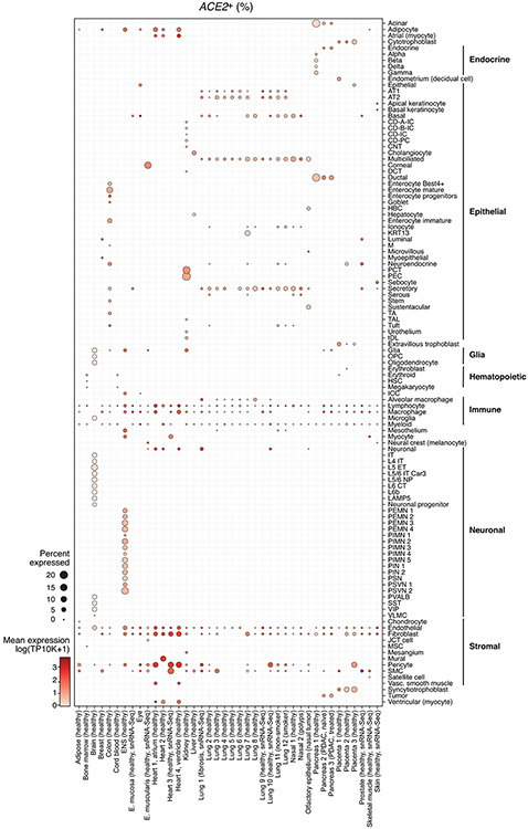
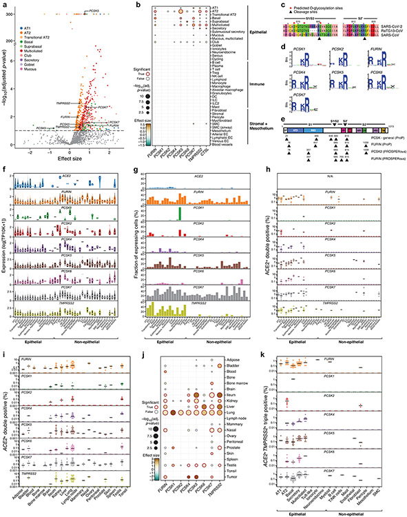
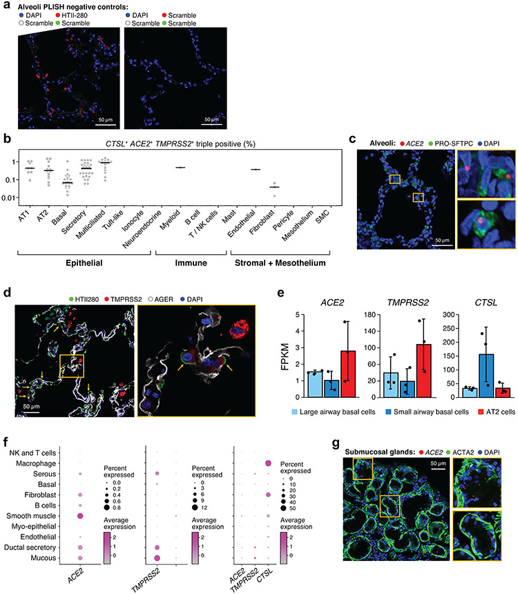
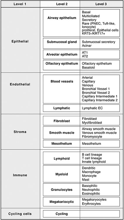
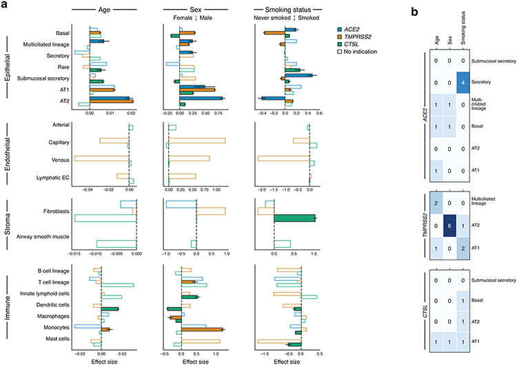
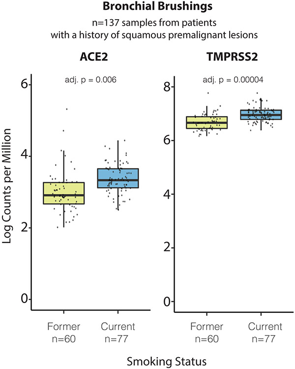
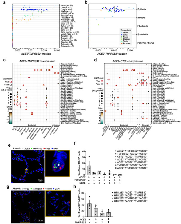
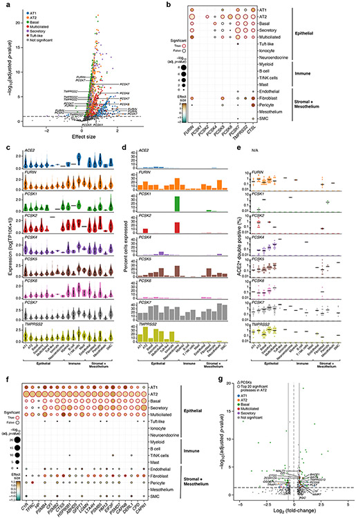
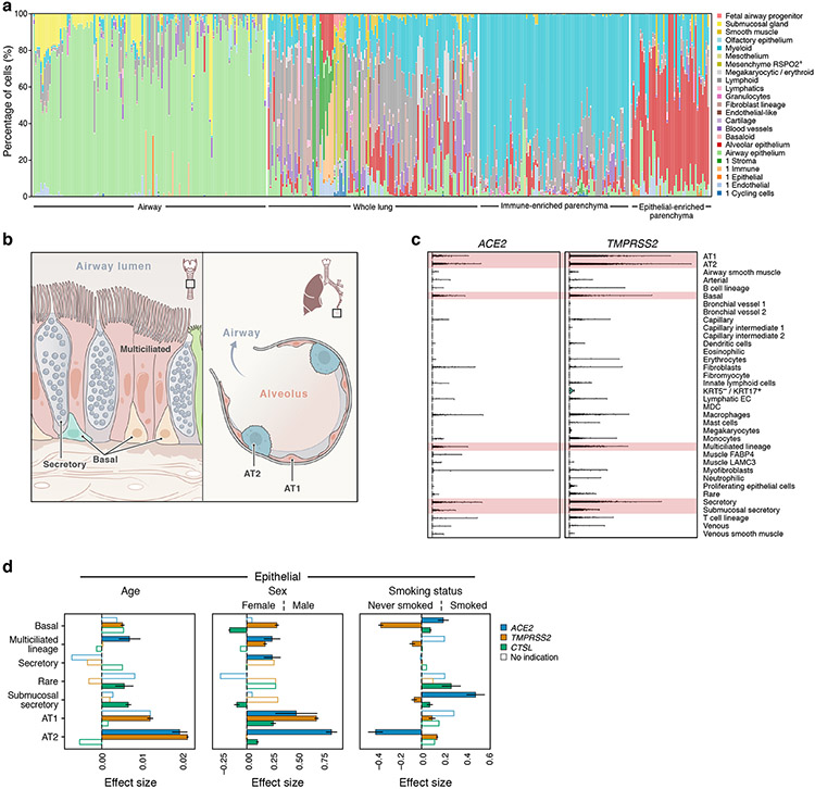
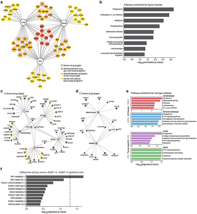
Angiotensin-converting enzyme 2 (ACE2) and accessory proteases (TMPRSS2 and CTSL) are needed for severe acute respiratory syndrome coronavirus 2 (SARS-CoV-2) cellular entry, and their expression may shed light on viral tropism and impact across the body. We assessed the cell-type-specific expression of ACE2, TMPRSS2 and CTSL across 107 single-cell RNA-sequencing studies from different tissues. ACE2, TMPRSS2 and CTSL are coexpressed in specific subsets of respiratory epithelial cells in the nasal passages, airways and alveoli, and in cells from other organs associated with coronavirus disease 2019 (COVID-19) transmission or pathology. We performed a meta-analysis of 31 lung single-cell RNA-sequencing studies with 1,320,896 cells from 377 nasal, airway and lung parenchyma samples from 228 individuals. This revealed cell-type-specific associations of age, sex and smoking with expression levels of ACE2, TMPRSS2 and CTSL. Expression of entry factors increased with age and in males, including in airway secretory cells and alveolar type 2 cells. Expression programs shared by ACE2TMPRSS2 cells in nasal, lung and gut tissues included genes that may mediate viral entry, key immune functions and epithelial-macrophage cross-talk, such as genes involved in the interleukin-6, interleukin-1, tumor necrosis factor and complement pathways. Cell-type-specific expression patterns may contribute to the pathogenesis of COVID-19, and our work highlights putative molecular pathways for therapeutic intervention.

摘要

血管紧张素转换酶 2(ACE2)和辅助蛋白酶(TMPRSS2 和 CTSL)是严重急性呼吸综合征冠状病毒 2(SARS-CoV-2)进入细胞所必需的,它们的表达可能揭示病毒的嗜性和对全身的影响。我们评估了 ACE2、TMPRSS2 和 CTSL 在来自不同组织的 107 个单细胞 RNA 测序研究中的细胞类型特异性表达。ACE2、TMPRSS2 和 CTSL 在鼻腔、气道和肺泡中的呼吸道上皮细胞的特定亚群中共同表达,也在与 2019 年冠状病毒病(COVID-19)传播或病理学相关的其他器官的细胞中表达。我们对 31 项肺单细胞 RNA 测序研究进行了荟萃分析,这些研究涉及来自 228 个人的 377 个鼻腔、气道和肺实质样本中的 1320896 个细胞。这揭示了 ACE2、TMPRSS2 和 CTSL 的表达水平与年龄、性别和吸烟之间的细胞类型特异性关联。进入因子的表达随着年龄的增长而增加,在男性中也是如此,包括气道分泌细胞和肺泡 2 型细胞。在鼻腔、肺和肠道组织中 ACE2TMPRSS2 细胞共享的表达程序包括可能介导病毒进入的基因、关键的免疫功能和上皮-巨噬细胞相互作用,例如涉及白细胞介素 6、白细胞介素 1、肿瘤坏死因子和补体途径的基因。细胞类型特异性表达模式可能有助于 COVID-19 的发病机制,我们的工作强调了潜在的分子途径,以进行治疗干预。

https://cdn.ncbi.nlm.nih.gov/pmc/blobs/5006/9469728/fa127bb2df53/nihms-1757283-f0005.jpg
https://cdn.ncbi.nlm.nih.gov/pmc/blobs/5006/9469728/c99583461add/nihms-1757283-f0006.jpg
https://cdn.ncbi.nlm.nih.gov/pmc/blobs/5006/9469728/679bc492c7d4/nihms-1757283-f0007.jpg
https://cdn.ncbi.nlm.nih.gov/pmc/blobs/5006/9469728/cbcf1c5c21d2/nihms-1757283-f0008.jpg
https://cdn.ncbi.nlm.nih.gov/pmc/blobs/5006/9469728/d1eff2cae95e/nihms-1757283-f0009.jpg
https://cdn.ncbi.nlm.nih.gov/pmc/blobs/5006/9469728/9d5fc4f2913e/nihms-1757283-f0010.jpg
https://cdn.ncbi.nlm.nih.gov/pmc/blobs/5006/9469728/ff1fd3c25ba9/nihms-1757283-f0011.jpg
https://cdn.ncbi.nlm.nih.gov/pmc/blobs/5006/9469728/32c6e6d7fe08/nihms-1757283-f0012.jpg
https://cdn.ncbi.nlm.nih.gov/pmc/blobs/5006/9469728/1489d846d32c/nihms-1757283-f0013.jpg
https://cdn.ncbi.nlm.nih.gov/pmc/blobs/5006/9469728/64ffd846a21b/nihms-1757283-f0014.jpg
https://cdn.ncbi.nlm.nih.gov/pmc/blobs/5006/9469728/a2ab3d80518b/nihms-1757283-f0015.jpg
https://cdn.ncbi.nlm.nih.gov/pmc/blobs/5006/9469728/598a817ae8de/nihms-1757283-f0001.jpg
https://cdn.ncbi.nlm.nih.gov/pmc/blobs/5006/9469728/0d6059108b5a/nihms-1757283-f0002.jpg
https://cdn.ncbi.nlm.nih.gov/pmc/blobs/5006/9469728/9e68ebb6f963/nihms-1757283-f0003.jpg
https://cdn.ncbi.nlm.nih.gov/pmc/blobs/5006/9469728/8a0d8512f306/nihms-1757283-f0004.jpg
https://cdn.ncbi.nlm.nih.gov/pmc/blobs/5006/9469728/fa127bb2df53/nihms-1757283-f0005.jpg