Zeng Jing, Cheng Qingqing, Zhang Dong, Fan Meng, Shi Changzheng, Luo Liangping
Medical Imaging Center, The First Affiliated Hospital of Jinan University, Guangzhou, China.
Engineering Research Center of Medical Imaging Artificial Intelligence for Precision Diagnosis and Treatment, Guangzhou, China.
Front Oncol. 2021 Feb 19;11:620628. doi: 10.3389/fonc.2021.620628. eCollection 2021.
Dynamic contrast-enhanced magnetic resonance imaging (DCE-MRI) now has been used to diagnose prostate cancer (PCa). Equivocal lesions are defined as PIRADS category 3 or a Likert scale of 1 to 5 category 3 lesions. Currently, there are no clear recommendations for the management of these lesions. This study aimed to estimate the diagnostic capacity of DCE-MRI for PCa and clinically significant prostate cancer (csPCa) in equivocal lesions.
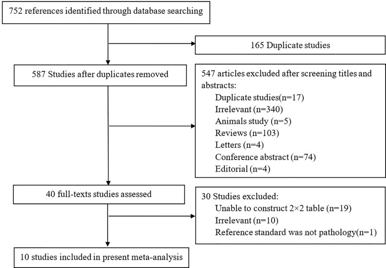
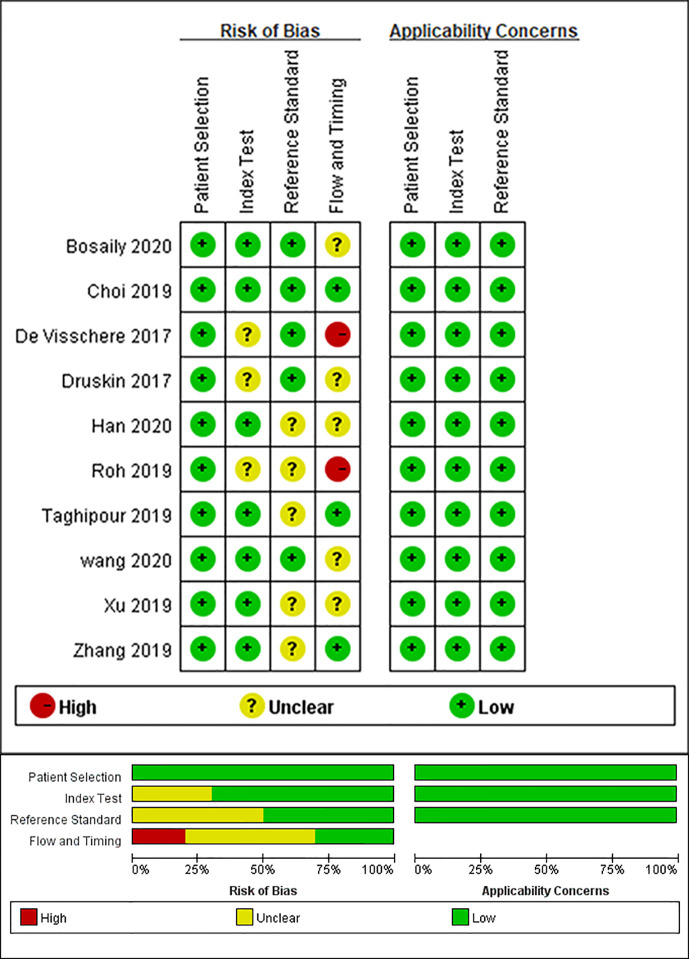
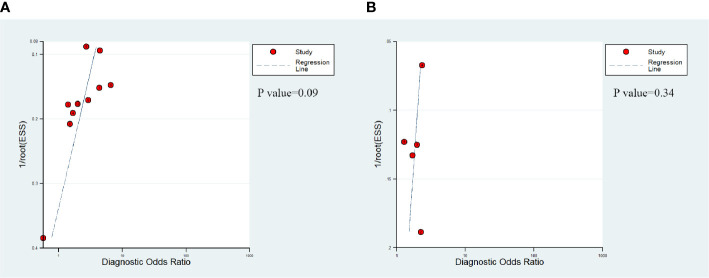
Two researchers searched PubMed, Embase and Web of Science to identify studies that met our subject. We searched for articles that mention the accuracy of the diagnosis of DCE-MRI for PCa or csPCa in equivocal lesions and used histopathological results as the reference standard. We used a tool (the Quality Assessment of Diagnostic Accuracy Studies-2 tool) to evaluate the quality of the studies that we screened out. Meta-regression was used to explore the reasons for heterogeneity in results.
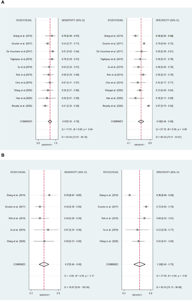
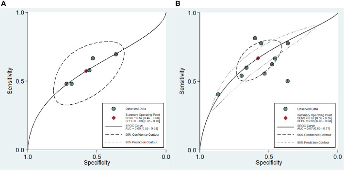
Ten articles were eventually included in our study. The sensitivity, specificity and 95% confidence intervals (CI) for DCE-MRI in diagnosing csPCa were 0.67 (95% CI, 0.56-0.76), 0.58 (95% CI, 0.46-0.68). The sensitivity and specificity and 95% CI for DCE-MRI in diagnosing PCa were 0.57 (95% CI, 0.46-0.68), 0.58 (95% CI, 0.45-0.70). The areas under the curve (AUC) of DCE-MRI were 0.67 (95% CI, 0.63-0.71) and 0.60 (95% CI, 0.55-0.64) while diagnosing csPCa and PCa. Through meta-regression, we found that study design, magnetic field strength, the definition of csPCa, and the scoring system were the sources of heterogeneity.
The results of our study indicate that the role of DCE-MRI in equivocal lesions may be limited.
动态对比增强磁共振成像(DCE-MRI)现已用于诊断前列腺癌(PCa)。可疑病变被定义为前列腺影像报告和数据系统(PIRADS)3类或李克特量表1至5级中的3类病变。目前,对于这些病变的处理尚无明确建议。本研究旨在评估DCE-MRI对可疑病变中PCa和临床显著前列腺癌(csPCa)的诊断能力。
两名研究人员检索了PubMed、Embase和科学网,以确定符合我们主题的研究。我们搜索了提及DCE-MRI对可疑病变中PCa或csPCa诊断准确性且以组织病理学结果作为参考标准的文章。我们使用一种工具(诊断准确性研究质量评估-2工具)来评估我们筛选出的研究的质量。采用Meta回归分析来探讨结果异质性的原因。
最终有10篇文章纳入我们的研究。DCE-MRI诊断csPCa的敏感性、特异性及95%置信区间(CI)分别为0.67(95%CI,0.56-0.76)、0.58(95%CI,0.46-0.68)。DCE-MRI诊断PCa的敏感性、特异性及95%CI分别为0.57(95%CI,0.46-0.68)、0.58(95%CI,0.45-0.70)。DCE-MRI在诊断csPCa和PCa时的曲线下面积(AUC)分别为0.67(95%CI,0.63-0.71)和0.60(95%CI,0.55-0.64)。通过Meta回归分析,我们发现研究设计、磁场强度、csPCa的定义及评分系统是异质性的来源。
我们的研究结果表明,DCE-MRI在可疑病变中的作用可能有限。